Pharynx Images: Key Learning Points for Residency
Aug 25, 2025
Nasopharynx
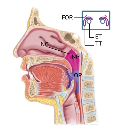
The nasopharynx lies behind the nasal cavity. The boundaries of the nasopharynx are as follows.
- Anteriorly, it communicates to the nasal cavity via choanae which has the nasal cavity anteriorly and the nasopharynx posteriorly. The choanae is separated in the midline by the vomer. The posterior ends of the interior and middle conchae from the lateral walls of the nasal cavity project medially into each choana.
- The lateral boundary is formed by an eustachian tubal opening which is covered by a cushion called torus tubarius. A recess is present behind this is called pharyngeal recess or fossa of Rosenmuller.
- The roof of the pharynx is formed by a basi-sphenoid which is continuous with the posterior wall.
- Posteriorly, there is the arch of Atlas with its overlying prevertebral cervical fascia and prevertebral musculature.
- The floor of the pharynx is formed by a soft palate. It is deficient posteriorly forming the nasopharyngeal isthmus which communicates with the oropharynx. The ridge separating the nasopharynx and oropharynx is called Passavant's ridge. It is formed by the fibers of the palatopharyngeus which constricts on swallowing to prevent regurgitation of food.
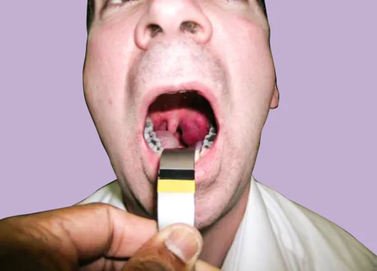
Oropharyngeal Isthmus
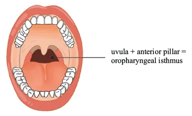
The oropharyngeal isthmus is the separation between the oral cavity and the oropharynx. It separates the oral cavity anteriorly from the oropharynx posteriorly. The anterior pillar is formed by a muscle called the palatoglossus muscle. The posterior pillar is formed by a muscle called the palatopharyngeus. Tonsils are present between palatoglossus and palatopharyngeus, called palatine tonsils. The oropharyngeal isthmus is formed by the uvula along with the anterior pillar.
Killian's Dehiscence
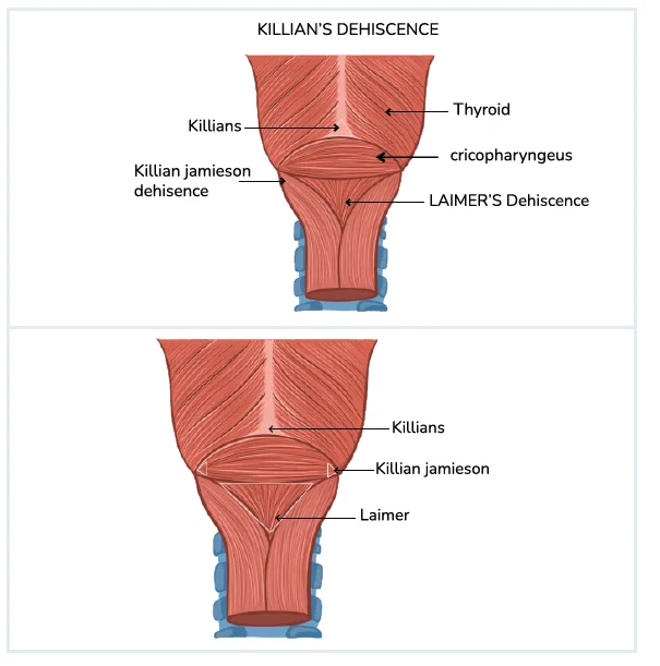
There are oblique fibers of the thyropharyngeus and horizontal fibers of the cricopharyngeus in the inferior constrictor. There is a triangular area of dehiscence called Killian's dehiscence.
Another dehiscence present laterally between the upper longitudinal fibers of the esophagus and the cricopharyngeus is called Killian's Jamison's dehiscence. The dehiscence in the midline between the cricopharyngeus and the longitudinal fibers of the esophagus is called Laimer dehiscence.
Cricopharyngeal Myotomy
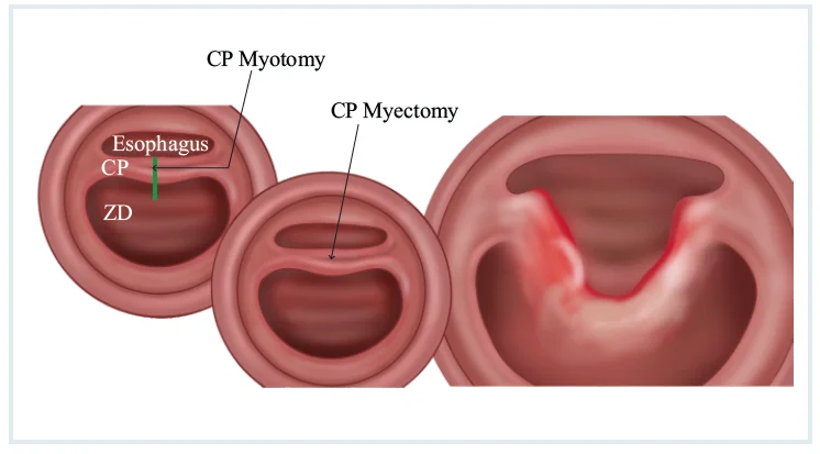
Pass a rigid esophagoscope up to the level of the cricopharyngeus muscle and rotate slightly to allow the light to shine through the skin and subcutaneous tissue. After identifying the cricopharyngeus muscle, do a cricopharyngeus myotomy, and the cricopharyngeus muscle is divided along the length for at least 3 cm.

Cleft Lip and Palate
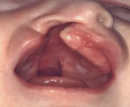
It is a common congenital abnormality associated with dysphagia. Deficiency of the lip is called cleft lip and deficiency of the palate is called cleft palate. Insufficient sucking occurs due to inadequate lip seal and inability to seal off the nasopharynx.
Plummer Vinson Syndrome
It is seen in middle-aged females. It has iron deficiency, genetic predisposition, and autoimmune. Factors. It is a premalignant condition and these patients have a higher possibility of developing squamous cell carcinoma.
Diffuse Esophageal Spasm
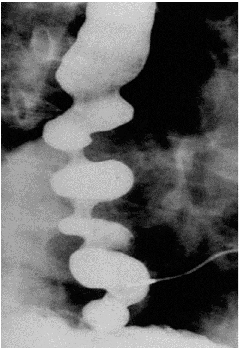
- More common in women, high emotional stress.
- Involves lower two thirds of esophagus.
- Hypertrophy of muscular layer of esophageal wall and degeneration of the esophageal branches of vagus nerve.
- Rapid wave progression down the esophagus.
- Dysphagia and chest pain.
- Corkscrew esophagus or pseudo diverticulosis.
Lymphoid Tissue Hyperplasia in The Pharynx
Lymphoid tissue hyperplasia in the pharynx commonly involves all the tissue of Waldeyer's ring including adenoidal, palatine and lingual tonsils. Adenoidal hyperplasia and hypertrophy may cause eustachian tube obstruction and otitis media with effusion. A biopsy of this tissue to exclude nasopharyngeal carcinoma and lymphoma is mandatory.
Thornwaldt's Cyst/Bursa
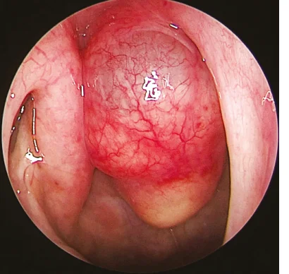
This is the most common epithelial growth next to adenoidal hypertrophy in the NP. The cyst is a result of a cleavage line between the nasal and pharyngeal embryological processes (Rathke's pouch). This lesion is usually asymptomatic although some patients may present with post-nasal drip due to the extrusion of the contents of the cyst occasionally. The diagnosis of the mass is usually incidental as part of a nasal endoscopic examination.
Paraganglioma
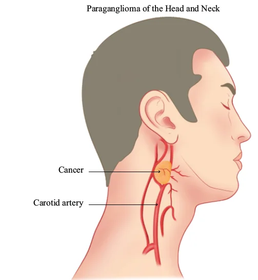
Paragangliomas are cells arising from the carotid bodies. These arise from paraganglionic cells, a component of the sympathetic nervous system.
Propel your ENT Residency Preparation! Access conceptual video lectures, QBank, and premium study resources on the PrepLadder App. Download it today!

PrepLadder Medical
Get access to all the essential resources required to ace your medical exam Preparation. Stay updated with the latest news and developments in the medical exam, improve your Medical Exam preparation, and turn your dreams into a reality!