Images in Otology: Key Learning Points for Residency
Aug 19, 2025
Image 1
Auricle
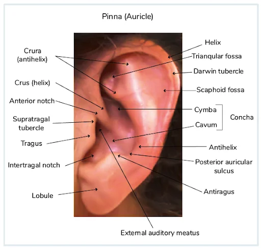
The pinna was actually developed during evolution from primitive beings to collect the environmental sound waves. The auricle or pinna projects from the side of the head to collect sound waves, and the meatus leads inwards from the auricle to conduct variations to the tympanic membrane. They are the first of a series of stimulus modifiers in the auditory apparatus. The pinna is structured as irregularly concave, faces slightly forwards, and displays numerous eminences and depressions.
Image 2
Otoscopic Examination
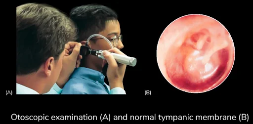
While performing an otoscopic examination:
In adults, the pinna needs to be pulled outwards, upwards, and backward, whereas in neonates downwards and backward. This maneuver can provide a clue to tenderness, which can indicate inflammation in the auricle and/or meatus.

Image 3
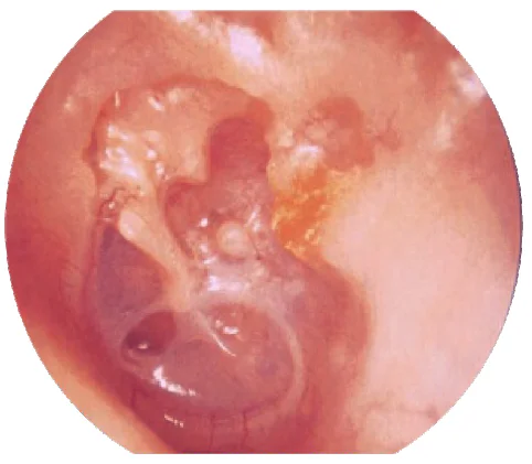
Prussak's Space
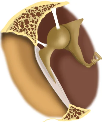
Prussak space is found between the pars flaccida and neck of the malleus , bounded by the lateral malleolar fold. this space can play an important role in the retention of keratin and subsequent development of cholesteatoma.
Image 4
Furuncle of the Right External Auditory Canal
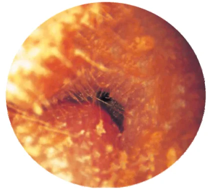
The endoscopic picture shows the cartilaginous section as there are visible hair follicles. Along with it, there is a projection and pus-filled like a pustular lesion in the external auditory canal.
Image 5
Technetium 99M Bone Scan
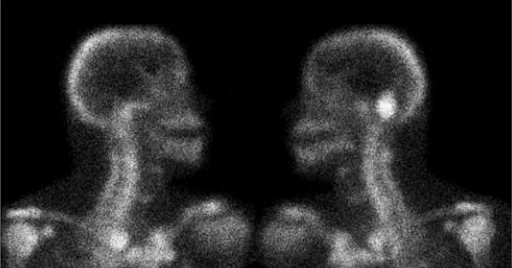
Shows areas of osteoblastic activity and is highly sensitive to bony infection. Traditional planar imaging or single photon emission computed tomography (SPECT) can be used. SPECT provides good anatomic localization and may highlight areas of bony involvement before the CT scan shows structural changes. Because bony repair persists long after the infection has resolved, it is not used to follow the response to treatment.
Image 6
Open Mastoid Cavities
The presence of open mastoid cavities should also be checked. The bony separation between the canal and mastoid is eroded. This is called an open mastoid cavity. It is usually created surgically. It can occur spontaneously by natural resolution of attic cholesteatoma. The resultant cavity is usually small and confined to the atticoantral region. These open mastoid cavities can be difficult to detect if there is a narrow external auditory meatus or the cavity is not in continuity with the attic or antrum.
Image 7
Sophono
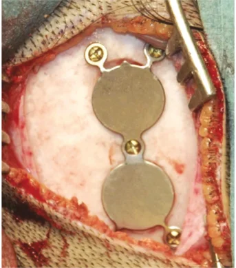
The Sophono system consists of an external sound processor coupled to a base plate with twin magnets. Internally, there are twin magnets implanted onto the bone without penetrating the overlying skin.
Image 8
Baha Attract System
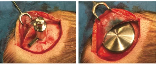
Image 9
Audiogram In Meniere's Disease
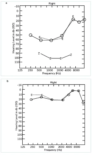
Hearing loss at low frequency is noted in Meniere's disease. After therapy for the disease, hearing loss comes back to normal.
Image 10
Brandt Daroff Exercises
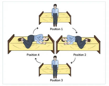
Mechanical self-treatment for p-BPPV. Should be performed for 15 minutes three times daily. This exercise consists of a rapid sequence of lateral head/body tilts. Starting from the sitting position, the patient rapidly moves to the challenging position, i.e., lying on the affected side (nose 45° up), and remains in this position for at least 30 seconds or until the vertigo subsides.
The patient then sits up for 30 seconds and then assumes the opposite head lateral and nose up position for 30 seconds before sitting up.
Download the PrepLadder app now to access high-yield content with 24-hr Free Trial. Explore premium study resources like Video Lectures, digital notes, QBank, and Mock Tests for a seamless exam preparation. Start your ENT Reasidency coaching journey with PrepLadder.

PrepLadder
Access all the necessary resources you need to succeed in your competitive exam preparation. Stay informed with the latest news and updates on the upcoming exam, enhance your exam preparation, and transform your dreams into a reality!
Navigate Quickly
Image 1
Auricle
Image 2
Otoscopic Examination
While performing an otoscopic examination:
Image 3
Prussak's Space
Image 4
Furuncle of the Right External Auditory Canal
Image 5
Technetium 99M Bone Scan
Image 6
Open Mastoid Cavities
Image 7
Sophono
Image 8
Baha Attract System
Image 9
Audiogram In Meniere's Disease
Image 10
Brandt Daroff Exercises