HMP Pathway Simplified: Role of NADPH in Metabolism & G6PD Deficiency Overview
Nov 7, 2025

HMP Shunt
It is a pathway where ATP is neither generated nor utilized. It acts as a source of NADPH. It acts as a source of Ribose 5-phosphate. Ribose 5 Phosphate is like a foundation on which every nucleotide is built. (Ribose 5 Phosphate is necessary for purine and pyrimidine nucleotide synthesis. Muscle Lacks Glucose 6 Phosphate Dehydrogenase. Hence, the oxidative phase is not operative in muscle.
Skeletal muscle has the gene (Glucose 6 Phosphate Dehydrogenase), but it's not active in skeletal muscle. And skeletal muscle does not require much NADPH. So, no oxidative phase happens in skeletal muscles. But skeletal muscle requires Ribose 5-phosphate. As a result of the reversible phase, 2 1⁄2 Glucose 6 Phosphate molecules it is reversed (as it is a reversible reaction), and Ribose 5 Phosphate is generated. Skeletal muscle Lacks Glucose 6 Phosphate Dehydrogenase, so the significance of the HMP Shunt in muscle is that it generates only Ribose 5 Phosphate in skeletal muscle.
|
|
NADH (Nicotinamide Adenine Dinucleotide Reduced)
NADH can enter the Electron Transport chain, ETC (ETC=2.5ATP), while NADPH cannot enter ETC & Cannot act as a source of ATP. NADH source is Glycolysis, Citric acid cycle, and Fatty Acid oxidation. All the paths which generate ATP, most of them will have NADH. NADPH is necessary as a coenzyme for the reductive biosynthesis of all lipids.
Lipids are more reduced than carbohydrates, and lipids give more energy than carbohydrates. (1g lipid = 9 calories while 1g of carbohydrates= 4 calories). The reason that lipids give more energy is that they are reduced. Anything that is reduced will give more energy. If lipids are reduced, it means synthesizing lipids; there will be multiple reduction steps. And for all steps hydrogen source is needed. And NADPH will act as a hydrogen source.
In Fatty acid synthesis, Cholesterol synthesis, Bile acid synthesis, etc. It is seen that NADPH is used as a coenzyme.
NADPH (Nicotinamide Adenine Dinucleotide Phosphate Reduced)
NADPH is necessary for regenerating GSH (an effective antioxidant in RBCs). Moreover, RBCs act as a major source of Oxidative stress in the body. RBCs also carry oxygen, in which there is a proficiency that oxygen is converted to superoxide radical, which initiates Oxidative stress. The major source of Oxidative stress in the body is RBCs. One of the antioxidant mechanisms that RBCs use is Glutathione (GSH).
NADPH is necessary as a coenzyme for Ribonucleotide Reductase. RNAs get converted to DNAs in the presence of Ribonucleotide Reductase, and for that hydrogen, a source is needed, NADPH. (RNAs – DNAs).
Steps of HMP Shunt: Two Phases
- Oxidative/Irreversible Phase: Acts as a source of NADPH.
This is initiated by Glucose 6 Phosphate Dehydrogenase (G6PD), which acts as Glucose 6 Phosphate. This is the first enzyme in the HMP Shunt. G6PD removes hydrogen and gives that hydrogen to its Coenzyme NADP, forming NADPH. And the Glucose 6 Phosphate becomes 6 Phosphogluconate. The next enzyme is 6 PGDH (Phosphate Gluconate Dehydrogenase).
Examples of enzymes that catalyze oxidative decarboxylation steps:
- PDH (Pyruvate Dehydrogenase) converts pyruvate to acetyl-CoA coA which catalyzes oxidative Decarboxylation steps.
- Alpha KGDH (Alpha-Ketoglutarate Dehydrogenase):
- ICDH (Isocitrate Dehydrogenase)
- 6PGDH (Phosphogluconate Dehydrogenase)
- BCKADH of Branchain Amino Acid Metabolism- The defect of the enzyme that causes MSUD (Maple Syrup Urine Disease).
Now, 6 Phosphogluconate Dehydrogenase catalyzes an oxidative decarboxylation step. So, hydrogen is removed from 6 Phosphogluconate and again given to NADP, forming NADPH. So for every glucose 6 Phosphate that gets into the HMP Shunt, 2 NADPH is formed. When it decarboxylates, CO² is removed. From a Hexose, because one carbon atom is removed, the product has to be a Pentose, and that Pentose is Ribulose 5 Phosphate. With this, the first phase is over. Both reactions of this phase are irreversible.
.jpg)
2. Non-Oxidative/ Reversible Phase: Acts as a source of Ribose 5-phosphate.
The Ribulose 5 Phosphate formed in the first phase needs to be converted to Ribose 5 Phosphate. The difference between Ribulose and Ribose is that Ribulose is a Ketose while Ribose is an Aldose. The next enzyme is Keto Isomerase, which converts Ribulose 5 Phosphate to Ribose 5 Phosphate.
Then Ribose 5 Phosphate will be used for purine and pyrimidine nucleotide synthesis. Not always Purine and Pyrimidine nucleotide synthesis. So excess or multiple Ribose 5 Phosphate would accumulate when there is no active Purine and Pyrimidine Nucleotide synthesis.
So these Ribose 5 Phosphate molecules will react with each other in the presence of Transketolase and Transaldolase (they go through a series of Transketolase and Transaldolase chemical reactions). And they try to regenerate all the Glucose 6 Phosphate back. But they won't be able to regenerate all the Glucose 6 Phosphate back, as there is the absence of carbon. Simultaneously, 3 Glucose and 6 Phosphate molecules react.
When 3 Glucose 6 Phosphate molecules react, initially, there would be 18 carbon atoms, and every Glucose 6 Phosphate will lose one carbon as Carbon Dioxide. So, from there, 3 Glucose and 6 Phosphate, three carbon atoms are lost. Then at the end, 15 Carbon atoms are left, which is 15C = 2½ Glucose 6 Phosphate. But half is not possible; it means whole. Therefore, the trio we get is G3P (Glyceraldehyde 3 Phosphate).
Transketolase- This enzyme needs Thiamine as its coenzyme. If there is a Thiamine deficiency, it means RBC Transketolase activity is low.
Simultaneously in HMP Shunt 3G6P —> 3CO2 + 2G6P + G3P + 6 NADPH + 6H+
HMP Shunt Defects
- Glucose 6 Phosphate Dehydrogenase Deficiency:
NADPH will not be generated when it has a defect. If NADPH is not generated, then GSH cannot be regenerated. So, without GSH, the antioxidant mechanism is missing in RBCs. RBCs become susceptible to lysis following exposure to oxidative stress.
- Hemolytic Anemia following exposure to oxidative stress:
In the absence of GSH, the antioxidant mechanism is missing in RBCs. RBCs become susceptible to lysis following exposure to oxidative stress. The history would be classical. An 11-year-old boy presented with hemolytic anemia after intake of primaquine (a prophylactic drug for malaria). Primaquine induces oxidative stress. So, until the person is very normal as per exposure to Oxidative stress because RBCs cannot regenerate Glutathione quickly, they become susceptible to Lysis following exposure to Oxidative stress ( for example: after intake of Primaquine or intake of Fava Bean).
Even normally, without consuming Fava bean or primaquine, it also causes Oxidative stress. So, there is a need for GSH to be regenerated in RBCs even normally, but that is met by two minor sources of NADPH: Cytoplasmic Isocitrate Dehydrogenase. Malic Enzymes will help in the regeneration of glutathione. Still, they have slowed up the pace, and when they get exposed to oxidative stress, there is a rapid rate at which hydrogen peroxide gets generated.
So, there should be a rapid pace at which Glutathione should be regenerated, and for this, a major NADPH source is needed, which is missing. So, they present with Hemolytic anemia following exposure to Oxidative stress.
- X-linked recessive:
5 defects which are X-linked-
- CHO (G6PD deficiency)
- Lipid metabolism (Fabry's disease, caused by Alpha Galactosidase, which is X-linked recessively inherited)
- Amino acid metabolism (Type 2 Hyperammonemia)
- Puri Salvage metabolism- GPRTase defect, which is an enzyme of the Purine Salvage pathway, and the Defect of this enzyme causes Lesch Nyhan Syndrome.
- MPS (Mucopolysaccharidosis)- Hunter's, caused by defects of Iduronate sulfatase.
Why should you provide a source of 2,3 BPG in stored blood?
It is because the stored blood in a bag does not have a continuous supply of glucose. And this would result in less glycolysis rate.
When the glycolysis rate is less, 2,3 BPG production is less, which is a by-product of glycolysis. When there is no 2,3 BPG, the affinity of hemoglobin for oxygen is high. RBCs' affinity for oxygen will be high, and when affinity for oxygen is high, these RBCs will unload oxygen to tissues. And tissues would not get oxygen (suffer from Hypoxia). So, the entire purpose of blood transfusion will not be served if there is no source of 2,3 BPG. That is why in all the stored blood, there should be a source of 2,3 BPG, and that is inosine monophosphate (IMP).
Inosine Monophosphate is Hypoxanthine + Ribose 5 Phosphate. So, if you add IMP to stored blood, it enters into RBCs, and within RBCs, IMP will be split into Hypoxanthine and Ribose 5 Phosphate. Then Ribose 5 Phosphate gets into the HMP Shunt.
As Ribose 5 Phosphate is an intermediate of the HMP Shunt. And as a result of HMP Shunt, we get 2½ Glucose 6 Phosphate, which is Glyceraldehyde 3 Phosphate. This G3P then becomes 1,3 BPG in the presence of Glyceraldehyde 3 Phosphate Dehydrogenase. Then 1,3 BPG becomes 2,3 BPG, which helps in unloading oxygen to tissues.
Important MCQS
Q1. The number of NADPH synthesized, CO liberated, and Glucose-6-Phosphate regenerated from 3 2 Glucose-6-Phosphate are
A. 3,3,3
B. 6,3,3
C. 6,3,2,5
D. 6,0,2,5
Answer: C. 6,3,2,5
Q2. The main function of the HMP Shunt in skeletal muscle is
A. Synthesis of NADPH
B. Synthesis of Ribose-5-Phosphate
C. Both
D. None
Answer: B. Synthesis of Ribose-5-Phosphate
Q3. Thiamine acts as a cofactor for all except
A. PDH
B. Isocitrate Dehydrogenase
C. Transketolase
D. a ketoglutarate Dehydrogenase
Answer: B. Isocitrate Dehydrogenase
Q4. Which of the following increases 2,3-BPG in stored blood:
A. IMP
B. Hypoxanthine
C. Phosphate
D. Citrate
Answer: A. IMP
A. Pyruvate kinase
Q5. Just before a planned departure to the tropics, a patient visits his physician complaining of weakness and noticing that his urine has recently become dark. Physical examination revealed a slightly jaundiced, yellow-colored sclera. Lab test indicated a low hematocrit, a high reticulocyte count, and significantly raised bilirubin. His MpMf was negative. His CKMM level was normal; he says this is his second visit, and he had some prophylactic measures during the previous visit. The probable enzyme Defect is.
A. Pyruvate kinase
B. Phosphofructokinase-1
C. Glucose-6-Phosphatase
D. Glucose-6-Phosphate Dehydrogenase
Answer: D. Glucose-6-Phosphate Dehydrogenase
Q6. Enzyme A and Enzyme B, respectively, are
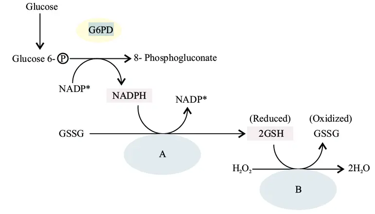
A. Glutathione Peroxidase Glutathione Reductase
B. Glutathione Reductase Glutathione Peroxidase
C. Glucose-6-Phosphate Dehydrogenase and 6-Phosphogluconate Dehydrogenase
D. 6-Phosphogluconate Dehydrogenase and Glucose-6-Phosphate Dehydrogenase
Answer: B. Glutathione Reductase Glutathione Peroxidase
Download the PrepLadder app now and unlock a 24-hour FREE trial of premium high-yield content. Access Smarter Video Lectures also in हिंglish, Game Changing Qbank, Audio QBank, Structured Notes, Treasures, Mock test for FREE to ace your NEET PG preparation. Elevate your study experience and gear up for success. Start your journey with PrepLadder today!

PrepLadder
Access all the necessary resources you need to succeed in your competitive exam preparation. Stay informed with the latest news and updates on the upcoming exam, enhance your exam preparation, and transform your dreams into a reality!
Navigate Quickly
HMP Shunt
NADH (Nicotinamide Adenine Dinucleotide Reduced)
NADPH (Nicotinamide Adenine Dinucleotide Phosphate Reduced)
Steps of HMP Shunt: Two Phases
2. Non-Oxidative/ Reversible Phase: Acts as a source of Ribose 5-phosphate.
HMP Shunt Defects
Why should you provide a source of 2,3 BPG in stored blood?
Important MCQS
Top searching words
The most popular search terms used by aspirants
- NEET PG Biochemistry
- NEET PG Biochemistry Preparation
PrepLadder Version X for NEET PG
Avail 24-Hr Free Trial