Rapid Revision Reignite Dermatology: Question-Answer Format
Sep 12, 2025


Bacterial Infections Of Skin
Big Question 1: Describe bacterial skin infections and their types.
Broad Answer: Bacterial skin infection known as pyoderma is further divided into follicular and non-follicular infections.
- Follicular infections
- Superficial folliculitis
- Deep folliculitis
- Furuncle
- Carbuncle
- Non-follicular infections
- Impetigo (most common in children, on the face).
- Types: Non-bullous Vs Bullous impetigo
- Erysipelas
- Acute Lymphangitis.
Detailed Questions
Q1.1: What is impetigo?
Answer:
- Impetigo is the most common bacterial infection, which is usually seen in children on the face.
- It is of two types:
- Non-bullous impetigo (Impetigo contagiosum) - more common
- Bullous impetigo
Q1.2: What is the difference between bullous and non-bullous impetigo?
Answer:
| Bullous impetigo | Non bullous impetigo /IC | |
| Age group | Usually seen in newborn | Usually seen in preschoolers/toddlers |
| Organism | Staph aureus | Both by strep and staph : Staph is more commonly seen in developed nations. Strep is more commonly seen in developing nation. If both are in option one should mark strep |
| C.F/type of crust | Varnish crust | Crusted erosions covered by honey colored crust. |
| Superficial bulla Sometimes filled and my gravity they appear half mood because the fluid in it settles down. Also called hypopyon | ||
| Complication | These patients may land up into Staphylococcal scalded skin syndrome (SSSS). | The common complication is Poststreptococcal glomerulonephritis (PSGN) |
Q1.3. What structure is affected in Bullous Impetigo (BI)?
Answer: BI-DSG-1 → The target, cleaved by the exfoliative toxin of Staphylococcus aureus.
Q1.4. What is the treatment of Impetigo?
Answer: Topical antibiotics such as Mupirocin and Fusidic acid are first-line.
Q1.5: What is the difference between erysipelas and cellulitis?
Answer:
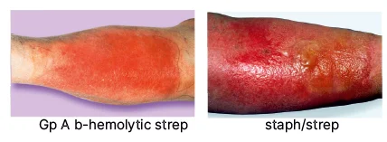
| Feature | Erysipelas | Cellulitis |
| Type | Superficial soft tissue infection | Deeper soft tissue infection |
| Site of involvement | Superficial dermis & lymphatics | Subcutaneous tissue |
| Lesion characteristics | Warm, red, indurated plaques | |
| Margins | Well-defined | Ill-defined |
| Systemic symptoms | Fever, constitutional symptoms | |
| Treatment | Oral antibiotics, leg elevation, NSAIDs | |
Cutaneous Tuberculosis And Leprosy
Big Question 2: Discuss the different forms of leprosy and their management.
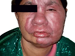
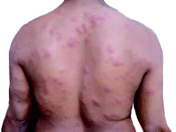
Broad Answer: Leprosy, caused by Mycobacterium leprae, is diagnosed by sensory loss in skin lesions, thickened nerves, and acid-fast bacilli on skin smear. It's classified by the Ridley-Jopling system (TT to LL) or WHO criteria (PB vs. MB). The Morphological Index (MI) tracks treatment response. Clinical features range from isolated lesions in TT to widespread nodules and Leonine facies in LL. Reactions (Type 1 & 2) are managed with steroids and continued MDT.
Detailed Questions
Q2.1: What are the three cardinal signs and diagnosis of leprosy?
Answer:
- Cardinal signs:
- Diminished or total loss of sensation in an atypical skin lesion.
- Enlargement and tenderness in a peripheral nerve.
- Finding of acid-fast bacilli on a slit skin smear.
- Detection of bacteria
- Slit skin - AFB
- Culture - cannot be cultured, can grow in a mouse foot pad or a nine-banded armadillo
- Serology
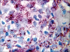
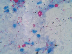
Q2.2: Explain the procedure for a slit skin smear.
Answer: A slit skin smear is performed by taking a 15 mm blade and making an incision 5 mm long and 3 mm deep - Rotate the blade and scrape out of the tissue - Smear the tissue and do ZN staining - Acid-fast bacilli appear pink on a blue background (do not get colorized). Identification criteria. Site: Ear lobe The density of bacilli in tissue: 10 bacilli/ gm
Q2.3: What are the types of bacteria?
Answer: 3 Types of bacilli
- Solid M. leprae: Living bacilli
- Fragmented: Dead bacilli
- Granular: Dead bacilli
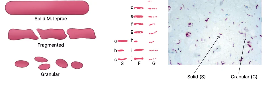
Q2.4: What is the Morphological Index (MI)?
Answer: It is the percentage of solid-stained (living) bacilli among 200 singly lying bacilli. The MI is the most useful index for monitoring treatment response and detecting drug resistance, unlike the Bacteriological Index (BI) which includes dead bacilli.
Q2.5: What is the Bacteriological Index (BI)?
Answer: Density or concentration of bacilli, living and dead bacilli ® Not useful in monitoring treatment.
| Grade | |
| 1-10 bacilli in 100 fields | 1+ |
| 1-10 bacilli in 10 fields | 2+ |
| 1-10 bacilli per field | 3+ |
| 10-100 bacilli per field | 4+ |
| 100-1000 bacilli per field | 5+ |
| >1000 bacilli/clumps/globi in every field | 6+ |
Q2.6: What are the two poles of leprosy?
Answer:
| Tuberculoid | Lepromatous |
| ● Here, the immune system is good, ● possible to curtail the bacilli. ● CMI or Cell-Mediated Immunity is protective against M.leprae of TH1 type. ● It has a TH1 response | ● Here, the immune system is not good, ● It is not possible to curtail the bacilli. ● The number of bacilli is more. ● Skin lesions will be more. ● It is more of a TH2 response. ● TH2 is protective of bacteria and not against an individual. ● There will be more antibodies. |
Big Question 3: Differentiate between Type 1 and Type 2 reactions.
Broad Answer:
| Type 1 | Type 2 | |
| Hypersensitivity reaction | Type 4 hypersensitivity reaction | Type 3 hypersensitivity reaction |
| Spectrum | Upper | The lower end of the spectrum |
| Predisposing factor | Happens after initiation of MDT | Stress, infection, and pregnancy bring down the immune system further. |
| Clinical feature | Exacerbation of existing lesions- swelling, redness, tender | New crops of lesions, along with the evening rise in temperature Constitutional + systemic symptoms |
| Nerve involvement | Tenderness, abscess formation | Not seen much here. |

Detailed Questions
Q3.1: What are the two types of leprosy reactions?
Answer: The two types are Type 1 reaction (or reversal reaction) and Type 2 reaction (or Erythema Nodosum Leprosum - ENL).
- Type 1 is a Type 4 hypersensitivity reaction (cell-mediated).
- Type 2 is a Type 3 hypersensitivity reaction (immune complex-mediated).
Q3.2: What are the nerve involvements in Type 1 and Type 2 reactions?
Answer: Nerve involvement is a prominent feature of Type 1 reactions, presenting as nerve tenderness and the potential for nerve abscess formation. Nerve involvement is not as common in Type 2 reactions.
Q3.3: What is the treatment for Type 1 and Type 2 leprosy reactions?
Answer: Management: For both reactions, MDT must be continued.
| Type 1 Reaction | Type 2 Reaction |
| ● Continue MDT + Oral steroids ● Mild: NSAID, aspirin ● Neuritis: Steroids ● Nerve abscess: Steroids > drain the abscessօ Alternative drugs: AZA, cyclosporine, MTX, HCQS | Oral Steroids and MDT ● Thalidomideօ Second-line treatment for type 2. օ Use: Resistant to steroids or cannot be given medicine. օ Multiple crops of new lesions are associated with fever - Arise in the evening and settle in 24 to 48 hours with PIH - Evanescent lesions. |
Sexually Transmitted Infections
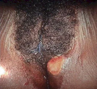
Big Question 4: What are the key types of genital ulcer diseases, and how can they be clinically differentiated based on their presentation?
Broad Answer: Genital ulcer diseases include Syphilis, Chancroid, Herpes genitalis, Donovanosis, and LGV. They differ in incubation period, number of ulcers, pain, base characteristics, lymph node involvement, and systemic symptoms. For example, syphilitic ulcers are single, painless, and indurated with bilateral painless lymphadenopathy. Proper identification is crucial for diagnosis and treatment.
Detailed Questions
Q4.1: What are the main types of Genital Ulcer Diseases?
Answer:
- Syphilis
- Chancroid
- Herpes genitalis
- Donovanosis
- LGV
Q4.2: How can different types of Genital Ulcer Diseases be differentiated based on clinical features?
Answer:
| Syphilis | Chancroid | HSV | Donovanosis | LGV | |
| Incubation period | 9-90 days | 2-7 days | 2-7 days | 3-30 days | 10-30 days |
| Ulcer number | One | Multiple | Multiple | One | Transient |
| Pain | No | Yes | Yes | No | No |
| Base | clean | Covered with greyish slough | Erosion | Granules | Nothing |
| Edges | Rubbery | Undermined | Polycyclic | Overhanging granules | Nothing |
| Induration | Positive | Negative | Negative | Ulcer may be firm | Nothing |
| Bleeds on touch | No | Yes | Yes | Yes very easily | Nothing |
| Lymph nodes | |||||
| Laterality | Bilateral | Unilateral | Bilateral | Pseudo Bubo | Bilateral |
| Pain | No | Yes | No | No | Yes |
Q4.3: What is the causative organism of Syphilis?
Answer: Treponema Pallidum
Q4.4: What happens if Syphilis is left untreated?
Answer: It can stay in the body for 10-40 years.
Q4.5: What is the incubation period of primary syphilis?
Answer: 10-90 days
Q4.6: How does a genital ulcer present in the primary stage of syphilis?
Answer: Chancre/ Hunterian chancre/ Hard chancre
If you’re looking to strengthen your final prep, don’t miss out on Rapid Revision Reignite in Question-Answer format by PrepLadder. It’s designed to help Medical PG aspirants cover the entire syllabus quickly with concise notes in a Question-Answer format, high-yield MCQs, and expert-led revision videos—perfect for last-minute reinforcement before the exam.
Download the PrepLadder app now to access high-yield content with 24-hr Free Trial. Explore premium study resources like Video Lectures also in हिंglish, digital notes, QBank, and Mock Tests for a seamless exam preparation. Time to begin your NEET PG coaching online with PrepLadder.

PrepLadder Medical
Get access to all the essential resources required to ace your medical exam Preparation. Stay updated with the latest news and developments in the medical exam, improve your Medical Exam preparation, and turn your dreams into a reality!
Navigate Quickly
Bacterial Infections Of Skin
Detailed Questions
Cutaneous Tuberculosis And Leprosy
Detailed Questions
Detailed Questions
Sexually Transmitted Infections
Detailed Questions
Top searching words
The most popular search terms used by aspirants
- NEET PG Dermatology
- NEET PG Dermatology Preparation