Anatomy of Middle Ear - NEET PG ENT
Feb 13, 2023

The anatomy of middle ear is a crucial topic for the NEET PG exam because the middle ear is a complex and vital part of the ear and auditory system. The anatomy of the middle ear is closely related to the physiology of hearing and balance.
A deep understanding of the anatomy of the middle ear helps in understanding and diagnosing conditions such as otitis media, tympanic membrane perforation, otosclerosis, and other ear diseases.
It is a critical aspect of otolaryngology, and a comprehensive understanding of its structure and function is essential for success in the NEET PG exam and in the practice of medicine.
Read this blog to get a quick overview of this important ENT topic.
MIDDLE EAR CLEFT
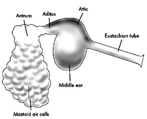
Middle ear cleft= Middle ear + Mastoid + ET
Middle ear volume is 1 ml. Mastoid volume is 5 ml. ET is closed at rest, so we don’t consider the volume. Middle ear cleft volume is 6 ml. Middle ear is communicating with the mastoid by an opening K/as Aditus. Mastoid antrum is the largest air cell in mastoid. Antrum is of adult size at birth
Middle ear mastoid is covered by a bony plate; that separates the middle ear mastoid from middle cranial fossa. The plate is K/as Tegmen or Dural plate. Tegmen /Dural plate is divided into 2 parts: The part that covers middle ear is K/as Tegmen tympani and the parts that covers mastoid is K/as Tegmen antri (just above antrum)
TM forms the lateral wall of the middle ear.

- TM divides middle ear into 3 parts:
- Part above TM is Epitympanum/ Attic
- Part In Front TM is Mesotympanum
- Part below TM is Hypotympanum
- Lateral bony wall of Epitympanum is K/as Scutum.
- Erosion of scutum is the important sign seen in CT scan of Primary Acquired Cholesteatoma. Roof of mastoid/middle ear is tegmen
- Tegmen has a bulge which can be seen from the cranial side. It is K/as Arcuate Eminence, it is due to push by superior semicircular canal. It is the important surgical landmark for facial nerve surgery in the middle cranial fossa approach.
Open Middle ear
- Anterior wall contains 2 openings
- Upper small opening for canal of Tensor tympani.
- Lower big opening for Eustachian tube/auditory tube (ET connects middle ear to nasopharynx)
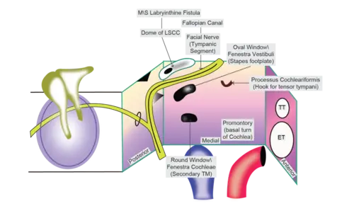
- Medial wall contains 2 windows
- Oval window/ Fenestra Vestibuli: It is covered by a footplate of stapes. Round window/Fenestra cochleae: It is covered by secondary TM.
- There is a hook-like structure K/as Processus Cochleariformis, which forms a hook for tensor tympani muscle. [Tensor tympani starts in the anterior wall from the canal and goes to medial where it turns at Processus Cochleariformis and comes out laterally and attaches to handle of malleus]
- Horizontal segment/tympanic segment of facial nerve. (Facial nerve is in the bony canal K/as fallopian canal, it is the largest bony canal for the CN, Length of fallopian canal is 27 mm) Postero superior to facial nerve there is a dome of lateral semicircular canal on medial wall of ME. This is the M/C site for Labyrinthine fistula
- There is a bulge in the medial wall K/as Promontory. It is due to the first turn/basal turn of cochlea.
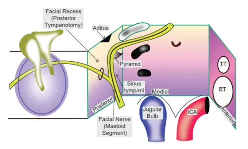
- Posterior wall
- Opening K/as Aditus which connects ME to mastoid. Vertical segment/ mastoid segment of facial nerve
- Pyramid: Stapedius muscle arises from here and attaches to the neck of stapes. Area medial to facial nerves is Sinus tympani (ME site for Recurrence / Residual cholesteatoma), it is a blind sac. Area lateral to facial nerve and above cauda facial angle is K/as Facial recess (this the M/C site for Posterior Tympanotomy)
- Inferior wall/Floor
- Floor/inferior wall
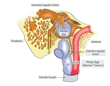
- Jugular bulb and internal carotid artery are present below the floor in between them there is a bony crest K/as Carotico Jugular Crest.
- Phelp sign: Inability to distinguish between Jugular bulb and Carotid artery due to erosion of carotico jugular crest seen in glomus tumor.
Also, read more medical notes blogs on different high-yield topics for strengthening NEET PG preparations. Download the PrepLadder app and get access to comprehensive study content curated by India’s best medical faculty.

PrepLadder Medical
Get access to all the essential resources required to ace your medical exam Preparation. Stay updated with the latest news and developments in the medical exam, improve your Medical Exam preparation, and turn your dreams into a reality!
Navigate Quickly
MIDDLE EAR CLEFT
MIDDLE EAR
Open Middle ear
PrepLadder Version X for NEET PG
Avail 24-Hr Free Trial