Helminthology : Structure, Classification, Growth, and Development
Apr 7, 2023

Helminthology, which is the study of parasitic worms, is an important topic in microbiology. Helminth infections are prevalent in many parts of the world, particularly in tropical and subtropical regions. Many helminth infections can cause serious illnesses and even death. As a medical professional, you must be able to diagnose and treat these infections appropriately to prevent severe outcomes.
| Cestodes | Trematodes | Nematodes |
| Tape like | Leaf like | Cylindrical |
| Monoecious (Both male and female in one) | Monoecious except schistosomes (Dioecious) | Dioecious |
| Suckers with hooks. | Suckers without hooks | Buccal capsule |
| Alimentary canal absent | Absent to poorly developed | Well developed |
| Body cavity absent | Absent | Present |
Let’s learn more about this important microbiology topic for NEET PG exam preparation.

CESTODES
|
Definitive Host |
Intermediate Host |
|
|
Man Man Dog Man Man |
Pig,Man Cattle Sheep / Man Man
|
Diphyllobothrium latum (Fish Tapeworm)
- Infective form → plerocercoid larvae
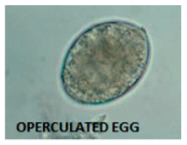
- Operculated egg (Lid)
- A/w megaloblastic anaemia
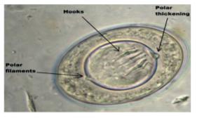
H. Nana (Dwarf Tapeworm)
- Infective form -Egg
- Egg has polar filaments, polar thickening with central hooklet
- Non-bile-stained egg
- Autoinfection - present
- DOC -Praziquantel
|
Important information Non-Bile-Stained Eggs
|
H. diminuta (Rat Tape worm)
- Autoinfection - Absent
- Egg lacks polar filaments
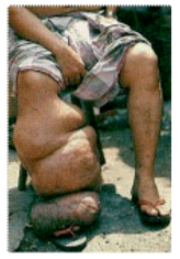
Echinococcus granulosus
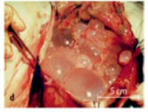
- Infective form – Eggs with contaminated food and water
- MC site – Liver (Hydatid cyst) >Lungs >Brain
Hydatid Cyst
- Most active layer – Endocyst (Germinal epithelium)
- It leads to formation of brood capsules, protoscolices and hydatid fluid.
- This can be detected by Casoni’s test (Type 1 HS reaction)
- Best treatment is surgical removal.
- PAIR therapy
Echinococcus Multilocularis
- Malignant Hydatid Disease
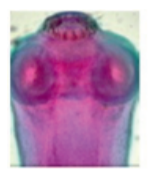
Tinea
T. Solium T. Saginata Longer Armed (Rostellum with Hooklets) Unarmed tapeworm as it lacks Rostellum and Hooklets Larval form A/K/a Cysticercus cellulosae Larval form A/K/a Cysticercus bovis Eating Uncooked pork leads to taeniasis (diarrhoea) Eating uncooked beef leads to taeniasis (diarrhoea) Infection form - Eggs majority of cases of Cysticercosis
Also, for Neuro- cysticercosis
MC site - Subarachnoid
Present with Atypical Seizures
IOC - CT / MRI (If calcified-CT)
DOC - AlbendazoleNo infection with eggs of taenia saginata

TREMATODES (LEAF LIKE FLUKES)

Monoecious
- 2 intermediate host
- 1st → Snail
- 2nd → Aquatic vegetation / Fish / Crab
- Infective form - Metacercariae (Cyst like structure)
- Operculated egg (lid) except Schistosomes
Schistosomes
- Dioecious
- IH → Snail
- Infective form → Cercariae
- Non operculated egg
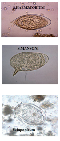
- S. Haematobium
- Resides in vesical plexus
- SCC of urinary bladder
- Terminal haematuria
- Egg has a Terminal spine
- S. Mansoni
- Resides in the inferior Mesenteric Plexus
- Katayama fever
- Swimmer’s itch (Cercarial dermatitis)
- Egg has a Lateral spine
- S. Japonicum
- Resides in Superior Mesenteric Plexus
- Katayama fever
- Eosinophilic Diarrhoea
- Egg has a Lateral knob / rudimentary spine
Fasciola Hepatica (Sheep Liver Fluke)
- Infection form – metacercariae
- 1st IH – Snail
- 2nd IH – aquatic vegetation. (Metacercariae) → (Man eats aquatic vegetation & develops infection)
- In bile duct, larvae mature to adult forms → Eggs are passed in faeces.
Clonorchis Sinensis (Oriental / Chinese Liver Fluke)
- 1st IH → Snail
- 2nd IH → Fresh water fish. (Metacercariae)
- Larvae matures in bile ducts to adult forms
- Constant irritation & inflammation of Bile duct → Carcinoma bile duct
Paragonimus Westermani (Lung Fluke)
- 1st IH → Snail
- 2nd IH → Crab / cray water fish (Metacercaria) → Rt lung → Erosions → Red brown sputum (Endemic haemoptysis)
Fasciolopsis Buski (Intestinal Fluke)
- Largest intestinal trematode
NEMATODES
Small Intestinal Large Intestinal Tissue Nematodes (Lymphatics) Subcutaneous Sites Ascaris Trichuris trichiura W.Bancrofti Loa Loa Ancylostoma Duodenale E.Vermicularis Brugia malayi Onchocerca volvulus Necator
AmericanusS.stercoralis
Infective Form
- By Eating Eggs
- E → Enterobius vermicularis
- A → Ascaris
- T → Trichuris trichiura
- By penetration of filariform larvae in sole of foot
- A → Ancylostoma duodenale
- N → Necator americanus
- S → Strongyloides stercoralis
- Go to lungs for maturation
- A → Ascaris
- A → Ancylostoma duodenale
- N → Necator americanus
- S → S. stercoralis
Large Intestinal Worms
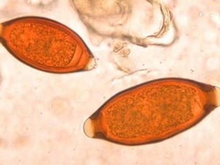
- T. Trichiura (Whipworm)
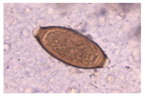
- Barrel shaped (dumbbell shaped) egg with mucus plugs.
- Bile stained (yellow)
- Rectal prolapse in children (coconut cake like rectum)
- Growth retardation
- A/W iron deficiency anaemia
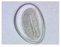
- Enterobius Vermicularis (Pin / Seat / Thread Worm Or Oxyuriasis)
- Perianal pruritus in children (MC)
- Autoinfection
- Egg - D shaped
- Has tadpole like larvae
- Plano convex egg
- Non bile stained
- NIH (National institute health) swab used
- Autoinfection
- Capillaria philippinensis, cryptosporidium parvum
- H. Nana
- E. Vermicularis
- S. Stercoralis
- Taenia Solium
Small Intestinal Worms
- Ancylostoma Duodenale (HookWorm)
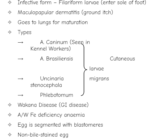
- Strongyloides Stercoralis
- Infection form – Filariform larvae
- Ovo – Viviparous
- A/W HIV +ve
- Parthenogenetic development →Female lays eggs without male.
- Autoinfection (Hyper infection syndrome in HIV +ve patients CNS and viscera are involved)
- Cutaneous larvae migrans (Larvae currens)
- Ascaris
- Infection form → Fertile Egg
- Lungs for maturation (2nd& 3rd moulting of larvae)
- A/W Intussusception
- Can l/t Intestinal obstruction
- Loffler’s syndrome (Massive eosinophilia)
- Female → Egg (Unfertilized egg)
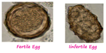
Fertile Egg Infertile Egg
- Eggs that Float on saturated salt solution
- Fertilised egg of ascaris
- A. Duodenale
- T. Trichiura
- E. Vermicularis
- H. Nana infertile Egg
Also Read:
TISSUE NEMATODES
W. Bancrofti

- Infective form – L3 filariform larvae
- Vector → Culex
- Viviparous (Microfilariae)
- Nocturnal periodicity (Come in blood at night)
- Sheathed (No nuclei in tail tip)
- Acute Filariasis
- Fever
- Lymphadenitis
- Lymphangitis
- Chronic Filariasis
- LN sclerosed and blocked so leads to
- Elephantiasis
- Hydrocele
- Chyluria
- Albuminuria
- LN sclerosed and blocked so leads to
- Granuloma Breast in females
- Lab Diagnosis
- Day time testing
- DEC given which irritates the larvae to come in blood at daytime
- Day time testing
- Blood smear examination done
- Sheathed microfilariae of W. bancrofti with no nuclei in tail tip seen
- Differentiated from Brugia malayi microfilariae is sheathed but has terminal 2 nuclei in the tail tip.
- IOC
- PCR for W. Bancrofti (Even picograms of DNA can be detected)
Subcutaneous Site Filariae
|
Skin |
Eye |
Fly (Transmitters) |
|
|
|
River blindness |
Simulium fly or black fly |
|
|
Loiasis |
Chrysops fly |
Trichinella Spiralis/ Muscle Worm
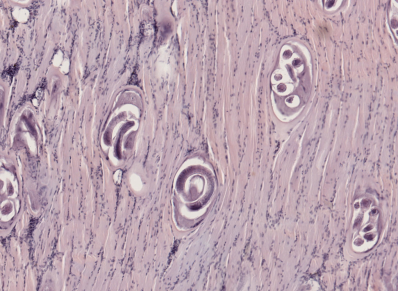
- Infective form → Larvae in uncooked pork
- Viviparous
Dracunculus Medinensis (Guinea Worm Disease)
- Eradicated in India (last case in Rajasthan)
|
Visceral Larvae Migrans |
Cutaneous Larvae Migrans |
|
|
Previous year Questions
Q. A child with 10 days abdominal pain presented to OPD. Stool microscopy was done which showed the given findings. What is the DOC for the disease caused by the given organism?
(AIIMS - May- 2018)
A. Albendazole
B. Mebendazole
C. Praziquantel
D. Pyrantel Pamoate
Q. Slow growing alveolar like tumour in liver?
(AIIMS - Nov - 2019)
A. E. Granulosus
B. E. Multilocularis
C. Cysticercus cellulosae
D. Amoebic liver abscess
Q. Which is correct about larval stage of Taenia solium?
(FMGE - June - 2019)
A. Larva currens
B. Cysticercosis Cellulose
C. Cutaneous Larvae Migrans
D. Visceral Larvae Migrans
Q. Which parasite lives in bladder plexus?
(FMGE - Nov - 2017)
A. Schistosoma
B. Fasciola
C. Ascaris
D. Echinococcus
Q. A 35-year-old man presented with dry cough & rusty coloured sputum. He has history of eating in Chinese restaurant very often with consumption of crabs often; what is the causative agent in this condition?
(NEET - Jan - 2019)
A. Diphyllobothrium latum
B. Pneumocystis jirovecii
C. Paragonimus Westermanii
D. Strongyloides stercoralis
Q. Identify the egg shown in image?
(FMGE - Aug - 2020)
A. Ascaris Lumbricoidoes
B. Enterobius Vermicularis
C. Trichuris Trichiura
D. H. Nana
Q. A 5-year-old child presented to OPD with complaints of rectal prolapse; on examination stunting & growth retardation was documented. What is parasitological cause for this clinical feature?
(NEET–Jan - 2019)
A. Trichuris Trichiura
B. Trichinella Spiralis
C. Giardia Lamblia
D. Enterobius vermicularis
Q. Identify the egg shown in image?
(FMGE - Dec - 2020)

A. Trichuris Trichiura
B. Enterobius Vermicularis
C. Ankylostoma
D. Ascaris
Q. Cutaneous larvae migrans caused by which organism?
(NEET–Jan - 2018)
A. Anisakiasis Simplex
B. Toxocara Species
C. Ancyclostoma braziliensis
D. Necator americanus
Q. Patients with history of kidney transplantation presenting with diarrhoea. The motility of the worms is given. Correct statement is?

(AIIMS - Nov - 2018)
A. Monoecious & the organism related with pathogenesis
B. Transmitted by intake contaminated food & water
C. Loffler’s pneumonia is not caused by same organism
D. Body gets this infection through filiform larvae
Q. Image shown here is?

(INICET - Nov - 2020)
A. Ascaris
B. Ancylostoma duodenale
C. Enterobius vermicularis
D. Strongyloides Stercoralis
Q. Pulmonary eosinophilia is seen d/t which of following infection?
(FMGE - Nov - 2017)
A. Ancyclostoma
B. Trichinella
C. Filaria
D. Roundworm
Q. A person working in which of the following profession can have the problem shown in the image?
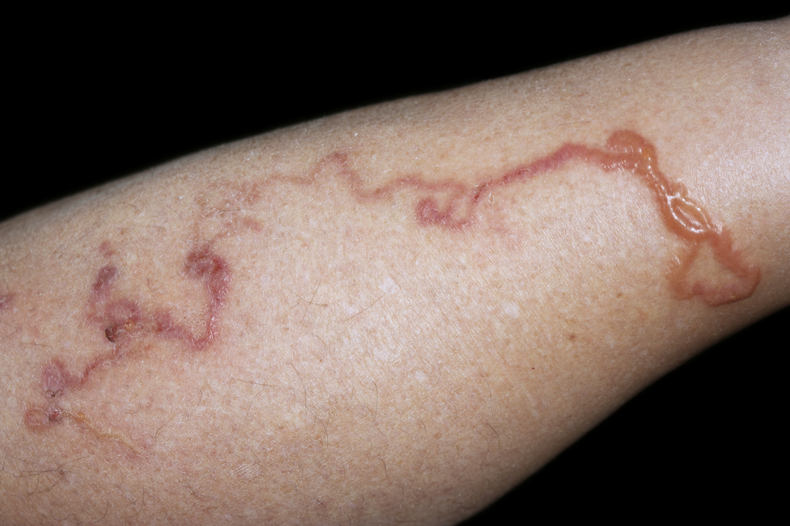
(AIIMS–May - 2019)
A. Person is working in the butcher house
B. Life guard in swimming pool
C. Kennel worker
D. Poultry farm worker
To study this high-yield microbiology topic in detail, download the PrepLadder app and find in-depth video lectures, study notes and MCQs covering the entire topic comprehensively.

PrepLadder Medical
Get access to all the essential resources required to ace your medical exam Preparation. Stay updated with the latest news and developments in the medical exam, improve your Medical Exam preparation, and turn your dreams into a reality!
Navigate Quickly
CESTODES
Diphyllobothrium latum (Fish Tapeworm)
H. Nana (Dwarf Tapeworm)
H. diminuta (Rat Tape worm)
Echinococcus granulosus
Hydatid Cyst
Echinococcus Multilocularis
Tinea
TREMATODES (LEAF LIKE FLUKES)
Monoecious
Schistosomes
Fasciola Hepatica (Sheep Liver Fluke)
Clonorchis Sinensis (Oriental / Chinese Liver Fluke)
Paragonimus Westermani (Lung Fluke)
Fasciolopsis Buski (Intestinal Fluke)
NEMATODES
Infective Form
Large Intestinal Worms
Small Intestinal Worms
TISSUE NEMATODES
W. Bancrofti
Subcutaneous Site Filariae
Trichinella Spiralis/ Muscle Worm
Dracunculus Medinensis (Guinea Worm Disease)
Previous year Questions
Q. A child with 10 days abdominal pain presented to OPD. Stool microscopy was done which showed the given findings. What is the DOC for the disease caused by the given organism?
Q. Slow growing alveolar like tumour in liver?
Q. Which is correct about larval stage of Taenia solium?
Q. Which parasite lives in bladder plexus?
Q. A 35-year-old man presented with dry cough & rusty coloured sputum. He has history of eating in Chinese restaurant very often with consumption of crabs often; what is the causative agent in this condition?
Q. Identify the egg shown in image?
Q. A 5-year-old child presented to OPD with complaints of rectal prolapse; on examination stunting & growth retardation was documented. What is parasitological cause for this clinical feature?
Q. Identify the egg shown in image?
Q. Cutaneous larvae migrans caused by which organism?
Q. Patients with history of kidney transplantation presenting with diarrhoea. The motility of the worms is given. Correct statement is?
Q. Image shown here is?
Q. Pulmonary eosinophilia is seen d/t which of following infection?
Q. A person working in which of the following profession can have the problem shown in the image?