Special X- ray Views in Orthopedics
May 2, 2025

45⁰ Lordotic View (Cephalic Tilt View)

Specific view for visualizing clavicle. The patient is in a standing position with a 45⁰ tilt.
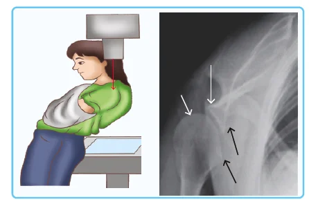
Serendipity View

- Also called Rockwood View
- Specific view for visualizing sternoclavicular joint
- The patient lies in a supine position
- Ray is passed at 45-60⁰
Zanca's View
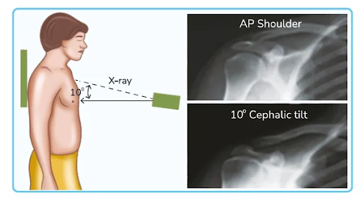
- It is a standing radiograph for the Acromioclavicular joint
- The X-ray beam is directed 10 to 15° Cephalad
- Demonstrates AC Joint & Distal Clavicle
- AC Joint Dislocation
- AC Joint Arthritis
- Distal Clavicular Osteolysis
- Measuring the Coracoclavicular Distance
WestPoint View

Specific view for demonstrating the Anterior aspect of the Glenoid Rim for detecting Bankart's lesion
The patient is prone on an X-ray table
↓
Abduct the affected arm away from the body for 90°
↓
The elbow flexed to allow the forearm to hang freely over the side of the table
↓
Rotate the head away from the affected side
↓
Place the cassette against the superior surface of the affected shoulder
.jpg)
Axial View

It is an appropriate projection to assess
- Suspected Shoulder Dislocations
- Proximal Humerus Pathology
- GlenoHumeral Articular Surface Abnormalities
Velpeau View
In Velpeau View, the beam is directed from superior to inferior through the shoulder with the patient's arm in a sling & with the patient leaning back. It is important to rule out Glenohumeral Dislocation.
Grashey's View

- The Shoulder AP Glenoid view is also known as a True AP or a 'Grashey's view.'
- Used to assess the
- The integrity of the Glenohumeral joint
- The articular surface of the Humerus
- Joint space for Subtle fractures such as a Bankart's lesion
- Post-Dislocation-Relocation,
- Proximal migration of Humerus
- Grashey's views (AP oblique) are obtained with the patient rotated 35-450, so the x-ray beam is parallel to the articular surface of the Glenoid.
Scapular Y View

The Scapular Y view is shot at an angle that separates the Scapula and Humerus from the ribs. It's essentially a true lateral of the scapula. The scapula looks like a Y when viewed laterally. Laterally, the body of the Scapula, Acromion, and Coracoid process converge at the Glenoid.
Stryker's Notch View

The Stryker notch view is a specialized projection of the shoulder aimed at assessing the Posterior Humerus. This is particularly useful in demonstrating
- Hill-Sachs deformities
- Gleno-Humeral Dislocation
Coyle's View

Also called the Trauma Oblique View of the Elbow, In the Coyle method, Sitting Axial Lateromedial projection, where the patient is seated, the elbow is flexed at 90°, palmar side downwards, with the X-ray tube at a 45° angle projection lateromedially.
Scaphoid View

The view is performed with the wrist in ulnar deviation to free the scaphoid from bony superimposition. Although performed PA, the view can be called an AP view.
Carpal Tunnel View

Clenched First View

The clenched fist view is an additional projection used to evaluate the suspected widening of the scapholunate interval, often performed bilaterally. It is a functional view that requires the patient to clench both hands. The signet ring sign in the scaphoid is indicative of scapholunate dissociation.
Brewerton's View

Metacarpophalangeal Joint flexed to 65° with the dorsum of the Proximal Phalanx flat against the radiograph cassette, and the beam angled 15° ulnar to radial profiles of the Collateral recesses. It is helpful to visualize collateral ligament Avulsion Fracture.
Danelius- Miller's View

An Axiolateral projection of the Acetabulum & the Proximal Femur to include the Head, Neck & Trochanters. This view is specifically done to visualize the Neck.
Robert's View

A True AP view of the Thumb taken with the Wrist maximal pronation with the dorsum of the thumb parallel to the table in which the beam is centered on the Trapezio-Metacarpal Joint. Used to assess Base of 1st Metacarpal fracture & Trapeziometacarpal arthritis. If instead of maximum pronation, the wrist is pronated to 30⁰, it is called Bett's view.
Dunn's View

It is a radiographic projection of the hip that demonstrates and examines the Hip joint, Femoral head, and Acetabulum, particularly the relationship between the Femoral Head and Acetabulum.
Positioning: Patient Supine, Hips flexed 90° & Abducted 20⁰. Beam-focused midpoint between Pubic symphysis & ASIS
Frog Leg Lateral View

In the Frog-leg view, the knee joint is flexed 30°-40° in a supine position, while the hip is externally rotated by 45° so that the image is taken toward the middle of the line connecting the upper Symphysis pubis and the Anterior- Superior Iliac Spine. Used to demonstrate DDH.
Clements Nakayama View

Modified Axiolateral View of the Hip. The Clements-Nakayama view of the Proximal. Femur is a highly specialized lateral projection utilized on patients with Bilateral Femoral. Fractures or patients unable to mobilize due to postoperative requirements.
Skyline Laurin or Axial View or Merchant View

The Knee Skyline Laurin view is an Inferior- Superior projection of the Patella. Knee is flexed to 45⁰.
Sunrise View

Here, the Patella appears to be rising over the horizon, The radiograph is taken with the X-ray beam tangential to the Patella parallel to the long axis of the lower extremity. In the Merchant view, the Knees are Bent at 45 degrees, and the muscles are relaxed, allowing the Patella to settle into the Trochlear groove, but in the sunrise view, the Knees are at Maximum Flexion (115⁰).
Download the PrepLadder app now and unlock a 24-hour FREE trial of premium high-yield content. Access Smarter Video Lectures also in हिंglish, Game Changing Qbank, Audio QBank, Structured Notes, Treasures, Mock test for FREE to ace your NEET PG preparation. Elevate your study experience and gear up for success. Start your journey with PrepLadder today!

PrepLadder Medical
Get access to all the essential resources required to ace your medical exam Preparation. Stay updated with the latest news and developments in the medical exam, improve your Medical Exam preparation, and turn your dreams into a reality!
Navigate Quickly
45⁰ Lordotic View (Cephalic Tilt View)
Serendipity View
Zanca's View
WestPoint View
Axial View
Velpeau View
Grashey's View
Scapular Y View
Stryker's Notch View
Coyle's View
Scaphoid View
Carpal Tunnel View
Clenched First View
Brewerton's View
Danelius- Miller's View
Robert's View
Dunn's View
Frog Leg Lateral View
Clements Nakayama View
Skyline Laurin or Axial View or Merchant View
Sunrise View
PrepLadder Version X for NEET PG
Avail 24-Hr Free Trial