Compare & Recall: High-Yield Physiology Tables for NEET PG
Aug 1, 2025


Table 1
Secondary Active Transport
Symport - Functioning of Sodium-Glucose Linked Transporter (SGLT)
Antiport - Functioning of Sodium-Calcium Exchanger


Table 2
Donnan’s Effect
|
Extracellular Fluid |
Intracellular Fluid |
|
|
Na+ K+ Ca2+ Mg2+ Cl– HCO3 Phosphates SO4 Glucose Amino acids |
142 mEq/L 4 mEq/L 2.4 mEq/L 1.2 mEq/L 103 mEq/L 24 mEq/L 4 mEq/L 1 mEq/L 90 mg/dl 30 mg/dl |
10 mEq/L 140 mEq/L 0.0001 mEq/L 58 mEq/L 4 mEq/L 10 mEq/L 75 mEq/L 2 mEq/L 0 to 20 mg/dl 200 mg/dl? |
|
Cholesterol Phospholipids Neutral fat |
0.5 gm/dl |
2 to 95 gm/dl |
|
PO2 POO2 pH Proteins |
35 mm Hg 46 mm Hg 7.4 2 gm/dl (5mEq/L) |
200 mm Hg? 80 mm Hg 7.0 16 gm/dl (40 mEq/L) |
Table 3
Temporal Summation and Spatial summation
Temporal summation
Spatial summation


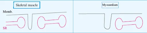
Table 4
Contraction Coupling of Skeletal Muscle and Cardiac Muscle
AP → Depolarization of T. tub. ↓ DHPR: activation ↓ DHPR interaction RyR1 (Mechanical) ↓ RyR opening ↓ Ca++ influx within cytoplasm ↓ Contraction AP → Depolarization ↓ Activation of DHPR ↓ Opening of DHPR ↓ ECF Ca++ enters into cell ↓ ⊕ RyR2 channel ↓ RyR2 opening ↓ Ca++ entry into cytoplasm ↓ Contraction
Skeletal muscle
Cardiac muscle
![]()
.jpg)
Table 5
Comparison Between Myocardium and Skeletal Muscles
Myocardium Muscles Skeletal Muscles Cells branch out with one another. These are single longitudinal fibres with no branching. Myocardium is a striated muscle. Skeletal muscle is a striated muscle. A single nucleus is present in each cell. Multiple nuclei are present in each cell. Intercalated discs are present. Intercalated discs are absent Gap junctions are present. Gap junctions are absent It is a functional syncytial tissue. It is not a functional syncytial tissue.
Table 6
Comparison of the SA Node and the AV Node
Sinoatrial Node (SA)
Atrioventricular Node (AV)
It is a sub-epicardial structure. Located close to Crista Terminalis.
It is located on the endocardial surface.
Dimensions: 10 mm long, 5 mm thick, and 1-2 mm deep
The AV node is compact (5x3x1 mm).
Physiological Location: Posterior wall of the right atrium near the superior vena cava opening.
Physiological Location: Base of the right atrial septum on the posterior aspect
Anatomical Location: Groove between crista terminalis and corresponding sulcus terminalis
Table 7
Generation of the 1st and Second Heart Sounds
First heart sound (1st HS) Second heart sound (2nd HS)
Table 8
Systolic and Diastolic Blood Pressure
Blood Pressure Component Determinant Relationship with Blood Pressure Systolic Blood Pressure Stroke Volume (SV) or Cardiac Output (CO) Directly proportional Compliance of the Aorta and Major Vessels Inversely proportional Diastolic Blood Pressure Elastic Recoiling Force of Aorta and Major Tissues Directly proportional Total Peripheral Resistance (TPR) Directly proportional
Table 9
Comparison Between the Epithelial Cells of PCT and Collecting Duct (CD)
Epithelial Cells of PCT Epithelial Cells of CD Brush border Present on the luminal side of the tube Absent Nature of tight junctions between the cells Leaky ' TJ' Tight 'TJ' Mode of transport of substances through the cells Paracellular transport: movement of small molecules, Na+, K+ through tight junctions.
Transcellular transport: movement facilitated by transmembrane channels and carrier proteins.No paracellular transport Only by transcellular transport. Carbonic anhydrase type 2
Present in the cytoplasmCarbonic anhydrase type 4 Present in the microvilli Absent


Table 10
Spasticity and Rigidity
|
Spasticity |
Rigidity |
|
|
|
|
|
|
|
|
Table 11
Physiological changes: NREM and REM
Aspect NREM REM Autonomic Outflow Parasympathetic outflow increases
Sympathetic outflow decreasesIncreased sympathetic outflow Cardiovascular Parameters Lower heart rate, lower BP Increase in heart rate Metabolic Rate Decreases - Temperature Decreases (brain and body) Decrease in brain and body temperature Muscle Tone and Reflexes Intact EMG: Silent, Muscle atonia (complete loss of muscle tone) Eye Movement (EOG) Absence of rapid eye movement Presence of rapid eye movement Electroencephalogram (EEG) Wave pattern not resembling wakefulness Beta-like wave pattern (Paradoxical sleep) Brain Electrical Activity Not associated with PGO (Ponto Geniculo Occipital) Associated with Ponto-Geniculo occipital (PGO) spike. PGO spike is proportionate to memory consolidation activity Sexual Responses No specific mention Penile erection (Parasympathetic activity), clitoral tumescence Dreaming Activity Not specified Active dreaming
Table 12
Difference between T4 & T 3
T4 T3 In secretion More (93% of total) Less (7% of total) Plasma conc. 8 μg/dL 0.15 μg/dL Protein-bound 99.98% 99.8% Free 0.02% 0.2% Free conc. 2 ng/dL 0.3 ng/dL Half-life (t1/2) 7 days 1 day Action Slower Faster Potency Less More (3-5 times) Binding to thyroid hormone receptors Low affinity High affinity
Download the PrepLadder app now and unlock a 24-hour FREE trial of premium high-yield content. Access Smarter Video Lectures also in हिंglish, Game Changing Qbank, Audio QBank, Structured Notes, Treasures, Mock test for FREE to ace your NEET PG preparation. Elevate your study experience and gear up for success. Start your journey with PrepLadder today!

PrepLadder
Access all the necessary resources you need to succeed in your competitive exam preparation. Stay informed with the latest news and updates on the upcoming exam, enhance your exam preparation, and transform your dreams into a reality!
Navigate Quickly
Table 1
Secondary Active Transport
Table 2
Donnan’s Effect
Table 3
Temporal Summation and Spatial summation
Table 4
Contraction Coupling of Skeletal Muscle and Cardiac Muscle
Table 5
Comparison Between Myocardium and Skeletal Muscles
Table 6
Comparison of the SA Node and the AV Node
Table 7
Generation of the 1st and Second Heart Sounds
Table 8
Systolic and Diastolic Blood Pressure
Table 9
Comparison Between the Epithelial Cells of PCT and Collecting Duct (CD)
Table 10
Spasticity and Rigidity
Table 11
Physiological changes: NREM and REM
Table 12
Difference between T4 & T 3