AP vs PA vs Lateral X-Ray: Differences, Positioning & When Each View Is Used
Sep 3, 2026
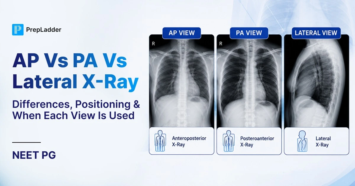
Chest radiography is the most frequently performed imaging study in clinical practice. Understanding the distinct positioning, technical limitations, and anatomical distortions of the Posteroanterior (PA), Anteroposterior (AP), and Lateral projections is essential for accurate diagnosis.
Misinterpreting an AP view as a PA view, for instance, can lead to a false diagnosis of cardiomegaly due to magnification artifact.
This guide provides a comprehensive breakdown of these three core radiographic projections, explaining how they are acquired, why they are used, and how to differentiate them on an examination.
Comparison: PA vs. AP Projections
| Feature | PA (Posteroanterior) View | AP (Anteroposterior) View |
| Beam Direction | Posterior to Anterior (Back to Front) | Anterior to Posterior (Front to Back) |
| Patient Position | Standing upright (Erect) | Supine or semi-erect (Bedside/Immobile) |
| Detector Position | Pressed against the anterior chest | Placed behind the patient's back |
| Source-to-Detector Distance | Long (~180-200 cm): minimal magnification | Shorter, variable (portable): more magnification |
| Heart Size | True to size (minimal magnification) | Artificially enlarged (magnification distortion) |
| Scapulae | Rotated laterally, clear of the lung fields | Not retracted; project over the lung fields |
| Clavicles | Angled downward (“BMX handlebar” position) | Sit higher, more horizontal (“mountain-bike handlebar” position) |
| Image Quality | Gold standard, high sharpness | Lower quality, often affected by poor inspiration/rotation |

Radiology Most Important 100 one-liners - Download Free PDF!
1. The PA (Posteroanterior) View
The PA view is the clinical “gold standard” for routine chest radiography.
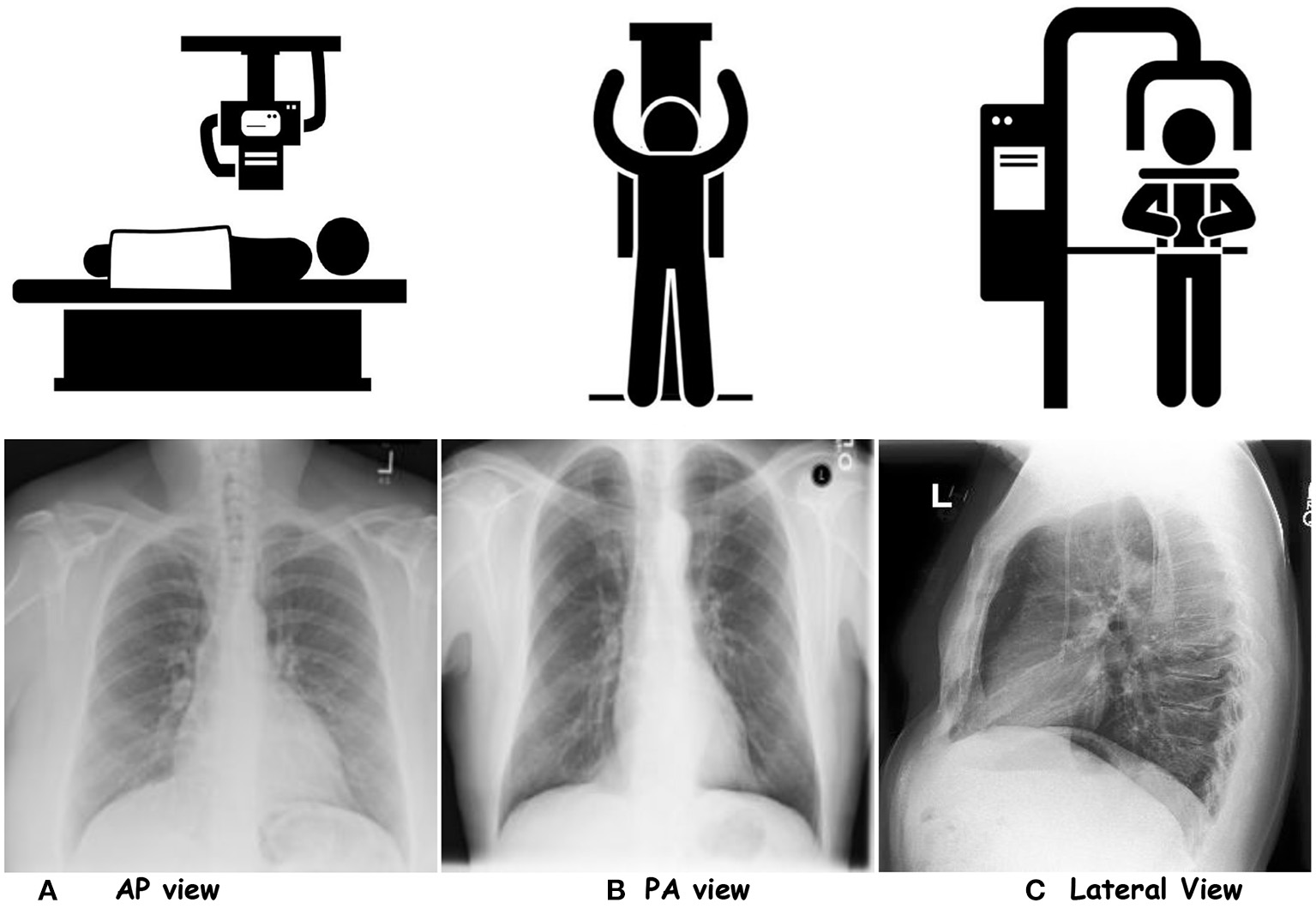
Positioning: The patient stands upright, facing the image receptor (detector) with their anterior chest pressed against it.
- The X-ray beam passes from the posterior (back) to the anterior (front).
Scapular Clearance: Patients are instructed to place their hands on their hips or hug the receptor side, which rotates the scapulae laterally so they do not obscure the lung parenchyma.
Cardiac Anatomy: Because the heart is an anterior mediastinal structure, placing the anterior chest against the detector puts the heart as close to the film as possible.
- This positioning, combined with a standard source-to-detector distance of roughly 180–200 cm (commonly cited as 6 feet), minimizes geometric magnification.
- A normal cardiothoracic ratio (<50%) can only be reliably assessed on a PA view.
Clinical Use: Used for all routine ambulatory evaluations to assess lung fields, pleural effusions, mediastinal contours, and true heart size.
2. The AP (Anteroposterior) View
The AP projection is a compromised view used when standard PA positioning is clinically impossible.
Positioning: The patient is usually supine (lying flat) or semi-erect in a hospital bed.
- The detector plate is placed behind their back, and the portable X-ray tube is positioned in front of their chest, typically at a shorter and more variable source-to-detector distance than a standard PA study.
Cardiac Magnification Artifact: Because the beam enters anteriorly, the heart is farther from the detector plate.
- The divergent X-ray beam projects a larger shadow onto the film, making the heart appear artificially enlarged (magnification distortion).
- Cardiomegaly cannot be definitively diagnosed on an AP film; however, if the heart appears normal-sized despite this magnification, true enlargement is unlikely, since magnification only makes the heart look larger, never smaller.
Anatomical Distortions: The scapulae are not retracted and usually project heavily over the lateral lung fields.
- The clavicles appear higher and more horizontal compared to the angled, downward appearance in a PA view.
- Furthermore, because the patient is often supine, pleural effusions may layer out posteriorly, creating a generalized hazy opacification rather than a distinct fluid meniscus.
Clinical Use: Reserved for critically ill, immobile, or trauma patients (e.g., in the intensive care unit, resuscitation room, or post-operative care) where bedside portable radiography is required.
- It is highly useful for checking the placement of medical devices such as endotracheal tubes, pacemakers, and central venous lines.
3. The Lateral View
A lateral chest radiograph is taken from the side, providing depth information that complements a frontal (PA) view.
Positioning: The standard is a “Left Lateral” projection, meaning the patient's left side is pressed against the detector.
- This minimizes cardiac magnification since the heart (predominantly left-sided structures) sits closest to the detector.
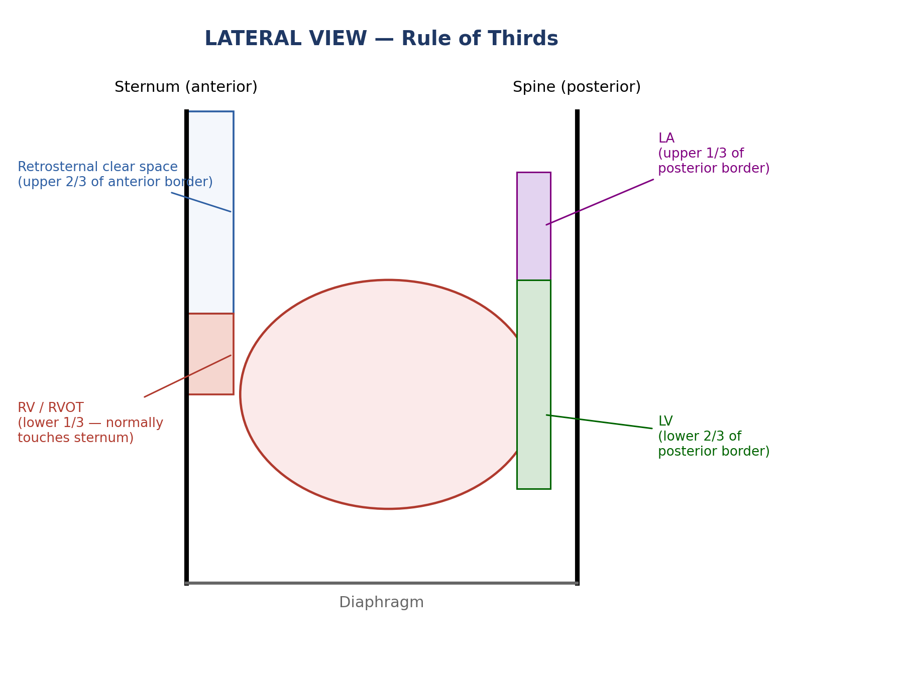
Retrosternal Clear Space: This is a crucial anatomical landmark, the normally dark (radiolucent) space directly behind the sternum, occupied by aerated lung.
- By the classic “rule of thirds,” the right ventricle (RV/RVOT) normally occupies only the lower one-third of the anterior cardiac border, touching the sternum, while the upper two-thirds should remain clear.
- Progressive filling of this space from below upward suggests right ventricular enlargement; opacity confined to the upper portion instead suggests an anterior mediastinal mass.
Posterior Cardiac Borders: The posterior border of the cardiac silhouette also follows a rule of thirds: the left atrium forms the upper one-third and the left ventricle forms the lower two-thirds.
- Posterior bulging beyond this line, formally assessed with Hoffman-Rigler's rule (measuring the distance between the posterior wall of the IVC and the posterior LV border, roughly 2 cm above the IVC–diaphragm junction), suggests left ventricular enlargement.
Clinical Use: Essential for locating lesions hidden behind the heart (retrocardiac space) or below the dome of the diaphragm.
- It is also highly sensitive for detecting small pleural effusions, as the posterior costophrenic sulcus is the deepest part of the pleural cavity and will show fluid blunting before the lateral sulcus seen on a frontal view.
Also Read: CT vs MRI in Brain Emergencies: When to Choose What | NEET PG 2026
How to Differentiate PA vs. AP on a Film
When presented with an unlabelled radiograph in an examination, use these rapid visual cues to determine the projection:
Look at the Scapulae:
- If the medial borders of the scapulae are clearly projected over the lung fields, it is highly likely an AP view.
- If they are pulled outward, clear of the lungs, it is a PA view.
Look at the Clavicles:
- AP clavicles sit high and horizontally across the lung apices.
- PA clavicles project downward in an angulated, V-shape over the lungs.
Look at the Gastric Bubble:
- An upright PA view will typically show a distinct gastric air-fluid level under the left hemidiaphragm, since an erect, horizontally-directed beam is needed to demonstrate an air-fluid interface.
- A supine AP view often lacks this distinct level, as gas distributes anteriorly across the stomach rather than layering out.
Also Read: Cephalohematoma: Types, Cases, Symptoms, Risk and Complications
Frequently Asked Questions
Why does the heart look bigger on an AP view?
This is caused by geometric magnification. The heart is an anterior structure. In an AP view, the detector is placed behind the back, increasing the physical distance between the heart and the film. As the X-ray beams travel from the front, they diverge, casting an exaggerated shadow of the heart onto the detector plate.
Can cardiomegaly be diagnosed on an AP film?
No. A cardiothoracic ratio greater than 50% on an AP view cannot reliably confirm cardiomegaly due to the magnification artifact. However, if the heart size appears strictly normal on an AP view, it is safe to conclude that it is not enlarged.
What structures form the cardiac borders on a PA view?
On a standard PA view, the right cardiac border is formed strictly by the right atrium. The left cardiac border is formed by the left ventricle and the left atrial appendage. The right ventricle is an anterior structure and does not form a distinct border on the PA view, which is why a lateral view is strictly required to assess right ventricular size.
What does it mean if the retrosternal or retrocardiac space is obliterated on a lateral view?
These are two distinct spaces and point to different problems. Filling of the retrosternal clear space (anterior to the heart, behind the sternum) suggests right ventricular enlargement or an anterior mediastinal mass.
Obliteration of the retrocardiac space (posterior to the heart, in front of the spine) suggests left ventricular enlargement or a posterior mediastinal/paraspinal mass; Hoffman-Rigler's rule can be used to formally confirm left ventricular enlargement in this view.
Understanding these precise positioning principles and anatomical landmarks ensures accurate diagnosis, appropriate image-quality assessment, and correct clinical interpretation of chest radiographs.

PrepLadder
Access all the necessary resources you need to succeed in your competitive exam preparation. Stay informed with the latest news and updates on the upcoming exam, enhance your exam preparation, and transform your dreams into a reality!
Navigate Quickly
Radiology Most Important 100 one-liners - Download Free PDF!
1. The PA (Posteroanterior) View
2. The AP (Anteroposterior) View
3. The Lateral View
How to Differentiate PA vs. AP on a Film
Frequently Asked Questions