Rapid Revision Reignite Radiology: Question-Answer Format
Sep 22, 2025

Cardiovascular System
Big Question 1: How are the heart borders and chambers identified on imaging, and what are the key radiological signs of chamber enlargement and mediastinal anatomy?
Broad Answer: On X-ray, the right heart border shows the right atrium; the left shows the left ventricle and aortic arch. On CT, the right ventricle is anterior, and the left atrium is posterior. LAE shows signs like the walking man and double density. Use 'SAAP' to identify mediastinal structures.
Detailed Questions
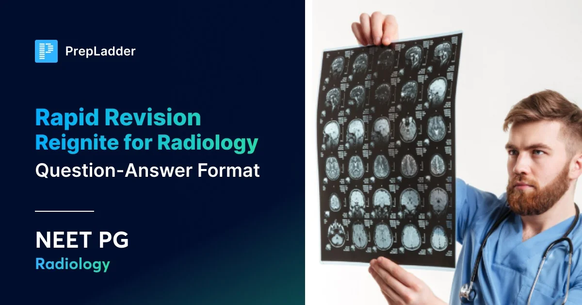
Q1.1: What anatomical structures form the right and left heart borders on a chest X-ray?
Answer:
| Heart Border | Structures Forming It |
| Right Heart Border | ● Right Atrium ● Superior Vena Cava ● Inferior Vena Cava ● Ascending Aorta (in the elderly) |
| Left Heart Border | ● Left Ventricle ● Left Auricle ● Pulmonary Artery ● Aortic Arch |
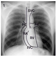
Right Heart Border
Left Heart Border
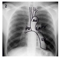
Q 1.2: What are the anatomical positions of the heart chambers as seen on a CT scan?
Answer:
- Anteriormost chamber of the heart: Right ventricle
- Posterior-most chamber of the heart: Left atrium
- Chamber On the left: Right atrium
- Chamber on the right: Left ventricle
Q 1.3: What are the key mediastinal structures seen on imaging?
Answer:
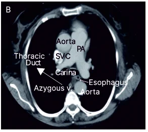
- From right to left: Mnemonic - 'SAAP'
- S- Superior vena cava
- AA- Ascending Aorta- anteriorly, Descending Aorta- posteriorly
- P- Pulmonary artery
Neuroradiology
Big Question 2: Describe the fundamental principles and key radiological findings of neuroradiology, focusing on the differentiation of imaging modalities and sequences.
Broad Answer:
- In neuroradiology, CT scans are identified by a "bone white" appearance.
- MRI sequences are distinguished by the appearance of water:
- T2-weighted
- T1-weighted
- FLAIR
- Advanced MRI techniques include DWI for acute infarcts (restricted diffusion), SWI for haemorrhages and calcification (blooming).
- MR Tractography for white matter tracts
- BOLD/fMRI for functional areas of the brain
- MR Spectroscopy, which provides a graph of metabolites to differentiate normal brain (NAA peak), tumours (Choline peak), and TB (Lipid peak).
- Myelography involves injecting contrast into the subarachnoid space to visualise the spinal cord.
Detailed Questions
Q2.1: How can a CT scan be differentiated from an MRI scan?
Answer:
- A CT scan shows a "bone white" appearance.
- An MRI scan shows a "bone cortex black."
Q2.2: How can you identify T1, T2, and FLAIR MRI sequences?
Answer:
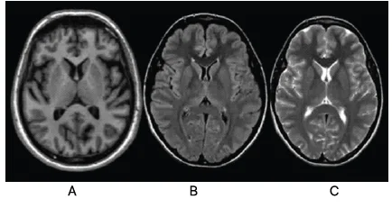
- T1-weighted MRI shows white matter as white and grey matter as grey. (Image A)
- T2-weighted MRI shows water as white. (Image C)
- FLAIR (Fluid Attenuated Inversion Recovery) shows all fluid, including CSF and white matter, as black. (Image B)
Q2.3: What is DWI (Diffusion Weighted Imaging) based on, and what does it show in an acute infarct?
Answer:
- ADC (Apparent diffusion coefficient) is based on the Brownian movement of protons (protons move freely like CSF ® Facilitated diffusion).
- An acute infarct shows restricted diffusion.
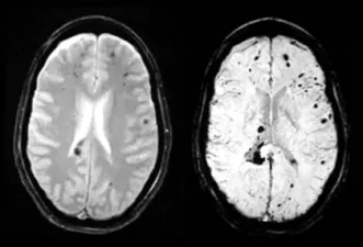
Q2.4: What is SWI (Susceptibility Weighted Imaging) used for, and how do haemorrhages and calcifications appear on it?
Answer:
- SWI is used for identifying haemorrhages and calcification based on magnetic susceptibility.
- These appear as black areas of blooming.
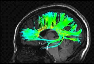
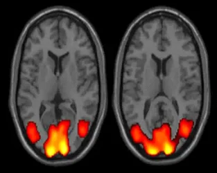
Q2.5: What is the purpose of MR Tractography/DTI and BOLD/fMRI?
Answer:
- MR Tractography/DTI is done to visualise white matter tracts and is based on the anisotropy of these tracts.
- BOLD/fMRI (Blood Oxygen Level-Dependent) is useful for locating the functional/eloquent areas of the brain (e.g., visual cortex, speech area) to aid in neurosurgery planning.
.jpg)
Respiratory Radiology
Big Question 3: Describe the radiological features of foreign body pathologies.
Broad Answer:
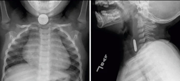
- Coin in Trachea vs Oesophagus
- Button Battery
- On AP view: Double ring appearance
- On lateral view: Sloping/ Bevelled edge
| Trachea | Esophagus |
| Appears as a SLIT in AP view Appears CIRCULAR in lateral view | Appears CIRCULAR in AP view Appears as a SLIT in lateral view |
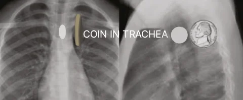
Coin in Trachea
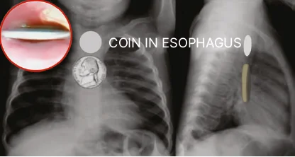
Coin in Esophagus
Detailed Questions
Q3.1: How can you differentiate a foreign body in the trachea from one in the oesophagus on an X-ray?
Answer: A foreign body in the trachea appears as a slit in the AP view and a circle in the lateral view. A foreign body in the oesophagus appears as a circle in the AP view and a slit in the lateral view.
Q3.2: What are the characteristic signs of a button battery on an X-ray, and what is its clinical significance?
Answer:
- On an AP view, it shows a double-ring appearance.
- On a lateral view, it shows a sloping/bevelled edge.
- A button battery is corrosive and must be removed even if the patient is asymptomatic.
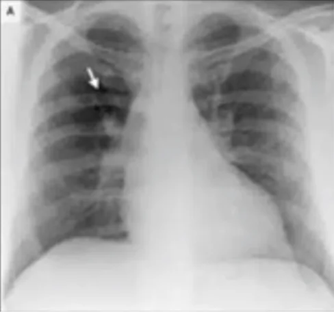
Q3.3: What is a "white out hemithorax," and what are its causes, with emphasis on the direction of tracheal shift?
Answer:
| Increased density | Decreased air | Fluid | |
| Seen in | Consolidation | Collapse Pneumonectomy | Pleural Effusion |
| Tracheal shift | No tracheal shift | Shift to the same side | Shift to the opposite side |
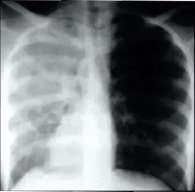
No tracheal shift

Shift to the same side
Shift to the opposite side
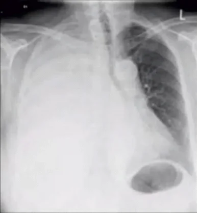
Q3.4: What is a "blackout hemithorax," and what conditions can cause it?
Answer:
| Increased air | Decreased density | Decreased vascular markings | |
| Seen in | Pneumothorax Emphysema | Post mastectomy Poland syndrome | Pulmonary embolism (Westermark sign) |
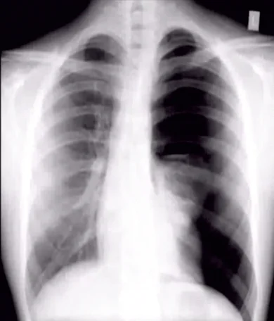
Pneumothorax Emphysema
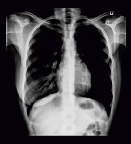
Post mastectomy Poland syndrome
Pulmonary embolism (Westermark sign)
Head and Neck
Big Question 4: What are the key radiological views and findings in Paranasal Sinus (PNS) and orbital imaging?
Broad Answer: Different X-ray views (like Water's, Caldwell's, Rhese, and Towne's) are used to visualize specific sinuses and orbital structures—e.g., Water's view for the maxillary sinus and These views for the optic canal. CT PNS, especially HRCT, is the investigation of choice before FESS.
Detailed Questions
Q 4.1: What are the different X-ray views used in PNS and orbital imaging, and which anatomical structures can be visualized in each?
Answer:
| View | Use |
| Caldwell's view (nose-forehead position; occipitofrontalview)- More elongated view | ● Superior orbital fissure ● Frontal sinus |
| Towne's view | ● Inferior orbital fissure |
| Rhese view | ● Optic canal ● Optic foramen |
| Water's view (nose-chin position; occipitomental view)- More rounded view ● Closed mouth ● Open mouth: Pierre's View | ● Maxillary sinus ● Floor of orbit |
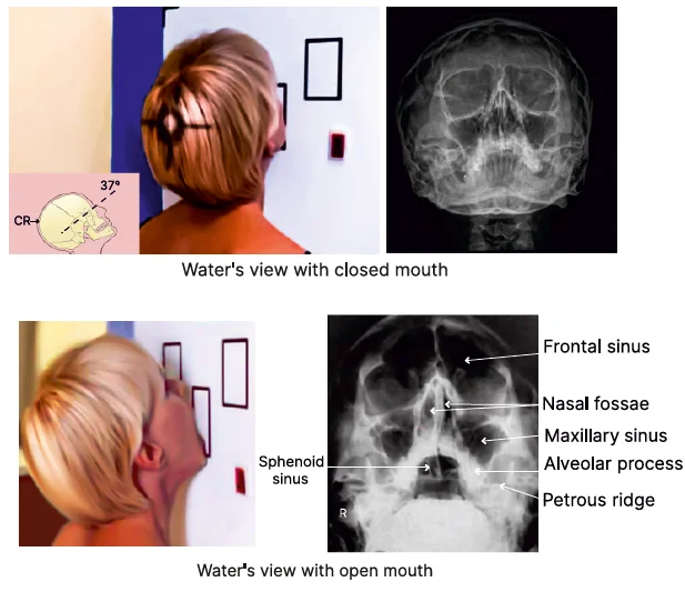
Q 4.2: In which X-ray views can the sphenoid sinus be visualized?
Answer:
● With an open mouth, the sphenoid sinus can also be visualized.
● Other views where the sphenoid sinus can be visualized: Basal view and Submentovertical view
Q 4.3: What is the investigation of choice before performing Functional Endoscopic Sinus Surgery (FESS)?
Answer: HRCT
Q 4.4: What is the HRCT finding of PNS?
Answer:
| 1. Concha bullosa (Pneumatization of middle turbinate) ● Concha- turbinate ● Bulla- air containing | 2. Onodi cell: Air cell related to optic nerve |
| 3. Haller cell: Infraorbital air cell | 4. Agger nasi: Anterior-most ethmoidal cell |
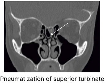
1. Concha bullosa (Pneumatization of middle turbinate)
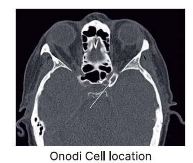
2. Onodi cell
3. Haller cell: Infraorbital air cell
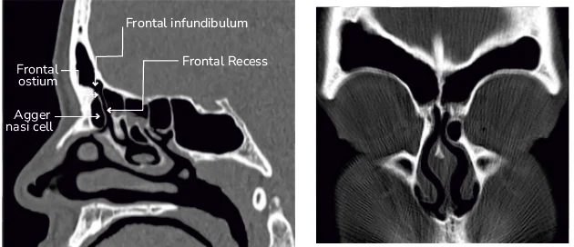
4. Agger nasi: Anterior-most ethmoidal cell
Also Read : NEET PG 2026: Important Topics For Radiology,
20 High Yield Radiology Flashcards NEET PG 2026
Gear up for your final prep with Rapid Revision Reignite by PrepLadder. Designed for Medical PG aspirants, it offers concise Q&A notes, high-yield MCQs, and expert-led revision videos—your perfect companion for last-minute success.
Download the PrepLadder app now to access high-yield content with 24-hr Free Trial. Explore premium study resources like Video Lectures also in हिंglish, digital notes, Audio QBank, and Mock Tests for a seamless exam preparation. Get started with NEET PG coaching online with PrepLadder.

PrepLadder Medical
Get access to all the essential resources required to ace your medical exam Preparation. Stay updated with the latest news and developments in the medical exam, improve your Medical Exam preparation, and turn your dreams into a reality!
Navigate Quickly
Cardiovascular System
Detailed Questions
Neuroradiology
Detailed Questions
Respiratory Radiology
Detailed Questions
Head and Neck
Detailed Questions
Top searching words
The most popular search terms used by aspirants
- NEET PG Preparation
- NEET PG Radiology
- NEET PG Radiology Preparation