Anesthesia Image Based Questions NEET PG
May 21, 2025

Q1. What is the mechanism of action of the given drug?

- Blocks Na+ channels
- Blocks K+ channels
- Both (1) & (2)
- Blocks Cl- channels
Ans. 1) Blocks Na+ channels
Explanation: Image shown is bupivacaine, a local anesthetic. All local anesthetic acts by inhibiting Na+ channels.
Q2. A 27-year-old male diagnosed with testicular torsion is scheduled for emergency surgery. During the pre-anesthesia assessment, the type of anaesthesia was decided. Which of the following about the needle (in the image below) used for administering the necessary anaesthesia is incorrect?

- It creates a smaller puncture
- Less chances of Post dural puncture headache
- Used commonly in spinal anaesthesia
- It is a dura-cutting needle
Ans. 4) It is a dura-cutting needle
Explanation:
- The image depicts a Whitacre needle, a dura-separating needle (not a dura-cutting needle) commonly used in spinal anaesthesia. (Option 3 ruled out)
- It has a conical, pencil-point tip with a small orifice, which is useful in reducing the incidence of Post dural puncture headache (PDPH)
Also read: NF-κB: Nuclear factor-κB Pathway defects
Q3. Refer to the readings depicted in the image. Which statement is inaccurate based on these readings?

- 76% is due to shivering
- 76% is due to Methemoglobinemia
- 76% is due to Carboxyhemoglobinemia
- 76% is due to Peripheral Vasoconstriction
Ans. 3) 76% is due to Carboxyhemoglobinemia
Explanation:
- The given image shows a pulse oximetry with a low SpO2.
- Out of 4 options, only carboxyhemoglobin can show false high readings, not false low readings.
Q4. Which of the following is the correct function of the shown image

- Monitor Intracardiac pressures
- Infuse solutions
- Measure Temperature
- All of the above
Ans. 4) All of the above
Explanation:
- The given image is of the Swan-Ganz catheter.
- These catheters monitor intracardiac pressures, and cardiac output, provide infusing solutions, and measure temperature.
Q5. Which of the following is an incorrect statement for the image below?

- The higher the value lower the awareness
- Elevated EEG is seen in Ketamine
- Depressed EEG is seen in COPD
- Nitrous oxide shows elevated EEG
Ans. 1) The higher the value lower the awareness
Explanation: The given image shown is of the bispectral index (BIS). Higher values on BIS indicate higher awareness
Also read: High-Yield One-Liners in Pediatric Immunology and Vaccines
Q6. Which of the following is the most common nerve-muscle combination tested?

- Ulnar Nerve & Adductor Pollicis Muscle
- Ulnar Nerve & Abductor Pollicis Muscle
- Radial Nerve & Adductor Pollicis Muscle
- Radial Nerve & Abductor Pollicis Muscle
Ans. 1) Ulnar Nerve & Adductor Pollicis Muscle
Explanation: The most commonly tested pair in neuromuscular testing is Ulnar Nerve & adductor pollicis muscle.
Q7. A 40-year-old female patient is undergoing a thyroidectomy under general anesthesia. A Train of Four (TOF) stimulation is performed to monitor neuromuscular function, the result of which is shown below. Which neuromuscular blocking agent was most likely administered?

- Succinylcholine
- Rocuronium
- Vecuronium
- Cisatracurium
Ans. 1) Succinylcholine
Explanation: The absence of fade with all twitches equally reduced indicates the use of a depolarizing neuromuscular blocking agent, such as Succinylcholine.
Q8. Which of the following is incorrect about the given image?

- D represents the best reflection of the alveolar CO2
- B to C represents CO2 of alveoli mixing with upper airway
- A to B represents Dead space ventilation
- E is the value seen on the monitor
Ans. 4) E is the value seen on the monitor
Explanation:
From Phase D to E
- Inhalation begins
- CO2 level drops to zero as oxygen fills the airway
- Point E does not represent the value seen on a capnograph monitor
- It is Point D, which represents the value seen on a monitor
Also read: Underlying Factors Behind Dementia
Q9. A 68-year-old man presents to the emergency department with palpitations, dizziness, and chest discomfort. His vital signs are blood pressure 75/50 mmHg, heart rate 190 beats per minute, and oxygen saturation 94% on room air. He appears pale and diaphoretic. ECG is shown below. What is the most appropriate next step in the management of this patient?

- Administer IV amiodarone 150 mg over 10 minutes
- Perform immediate unsynchronized defibrillation
- Perform immediate synchronized cardioversion
- Administer intravenous adenosine 6 mg
Ans. 3) Perform immediate synchronized cardioversion
Explanation:
- The clinical presentation and ECG suggesting a monomorphic wide-complex tachycardia with a regular rhythm is consistent with a diagnosis of an unstable monomorphic ventricular tachycardia.
- The first line of treatment for unstable VT with a pulse is synchronized cardioversion.
Q10. A 35-year-old woman presents to the emergency department with palpitations that started suddenly 30 minutes ago. She reports feeling lightheaded but denies chest pain or shortness of breath. Her vital signs show a heart rate of 180 beats per minute, blood pressure of 120/80 mmHg, and oxygen saturation of 96% on room air. ECG is shown below. Reveals a regular narrow-complex tachycardia without discernible P waves. What is the most appropriate initial management?

- Immediate synchronized cardioversion
- Intravenous adenosine 6 mg push
- Intravenous amiodarone 150 mg over 10 minutes
- Perform carotid sinus massage
Ans. 4) Perform carotid sinus massage
Explanation: The patient is likely experiencing stable supraventricular tachycardia (SVT). Initial management for stable SVT includes vagal maneuvers such as carotid sinus massage or the Valsalva maneuver.
Q11. A 65-year-old man with a history of coronary artery disease and heart failure presents to the emergency department after experiencing lightheadedness and near syncope. On examination, he is alert but pale, with a heart rate of 43 beats per minute and a blood pressure of 80/50 mmHg. The ECG is shown below. He remains hypotensive and symptomatic despite the IV administration of a maximum dose of atropine. What is the most appropriate next step in the management of this patient?

- Epinephrine IV bolus
- Amiodarone IV infusion
- Dopamine IV infusion
- Digoxin IV bolus
Ans. 3) Dopamine IV infusion
Explanation:
- The clinical presentation and ECG suggesting sinus bradycardia without evidence of heart block are consistent with symptomatic sinus bradycardia with hypotension.
- After atropine fails, the next step in the management is dopamine infusion.
Also read: Last 5 Last 5 Years PYQ's in Anesthesia for NEET PGYears PYQ's in Anesthesia for NEET PG
Q12. A 70-year-old man with a history of ischemic cardiomyopathy (ejection fraction 25%) and a previous history of myocardial infarction presents to the emergency department with dizziness, palpitations, and mild breathlessness. On arrival, his heart rate is 130 beats per minute, and his blood pressure is 100/64 mmHg. His ECG is shown below. Which among the following options is the most appropriate in the management of this patient?
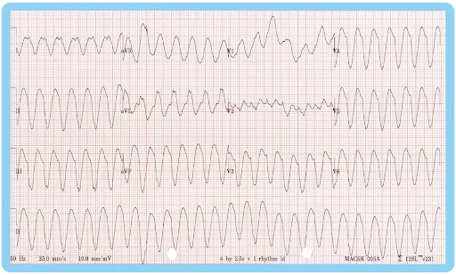
- Intravenous amiodarone 150 mg over 10 minutes
- Adenosine 12 mg IV push
- Intravenous lidocaine 1 mg/kg
- Intravenous magnesium sulfate 2 g
Ans. 1) Intravenous amiodarone 150 mg over 10 minutes
Explanation:
- The clinical presentation and ECG are suggestive of a stable, monomorphic wide-complex tachycardia, consistent with ventricular tachycardia (VT).
- Amiodarone 150 mg IV over 10 minutes, followed by 1 mg/min infusion for 6 hours, is the antiarrhythmic of choice for hemodynamically stable monomorphic ventricular tachycardia (VT) with impaired cardiac function (EF < 45%).
Q13. A 54-year-old woman presents to the emergency department with palpitations, dizziness, and mild shortness of breath. Her vital signs are a heart rate of 150 bpm, a blood pressure of 124/80 mmHg, and oxygen saturation of 95% on room air. The rhythm strip of the ECG is shown below. Vagal maneuvers are attempted without success. You decide to administer adenosine. Which of the following is the most appropriate initial dose of adenosine, and what is the mechanism of its action?

- 6 mg IV push, briefly depresses sinus node and AV node conduction
- 12 mg IV push, briefly depresses sinus node and AV node conduction
- 6 mg IV push, increases AV node conduction and depresses sinus node
- 12 mg IV push, decreases AV node conduction and increases sinus node rate
Ans. 1) 6 mg IV push, briefly depresses sinus node and AV node conduction
Explanation:
- The clinical presentation and ECG are suggestive of stable, narrow-complex regular tachycardia consistent with supraventricular tachycardia (SVT).
- Adenosine is an endogenous purine nucleoside that temporarily depresses the sinus node and AV node function, essential in treating regular, narrow-complex tachycardias like SVT. The initial dose of 6 mg IV should be administered rapidly, followed by a 20 mL saline flush.
Q14. Which of the following statements is/are incorrect regarding this type of euthanasia?

Statement 1: It is an act of commission
Statement 2: Not using extraordinary life-sustaining measures
Statement 3: It is a part of Hospice care
Statement 4: Practiced in India
- Statements 1 and 2
- Statements 2, 3 and 4
- Statements 3 and 4
- Statement 4 only
Ans. 2) Statements 2, 3 and 4
Explanation:
- The given image shows active euthanasia (as it shows the active administration of medicines).
- Statement 1 is correct, and Statements 2, 3, and 4 are incorrect.
Also read: Drugs included in Local Anaesthetics
Q15. Identify the method of assessment of Pain as depicted in the image.

- Wong-Baker Scale
- Visual Analog Scale
- CHEOPS
- McGill Pain Scale
Ans. 1) Wong-Baker Scale
Explanation:
- The given image is of Wong Baker’s Faces Scale
- Facial expressions are used to analyze the severity of pain.
Download the PrepLadder App and get the best NEET PG online coaching with world-class video lectures also in हिंglish, QBank, Mock Tests and more!
Download PrepLadder's best app for neet pg preparation for Android
Download PrepLadder's best app for neet pg preparation for Ios

PrepLadder Medical
Get access to all the essential resources required to ace your medical exam Preparation. Stay updated with the latest news and developments in the medical exam, improve your Medical Exam preparation, and turn your dreams into a reality!
Top searching words
The most popular search terms used by aspirants
- NEET PG Anesthesia
- NEET PG Anesthesia Preparation