Diaphragm: Development and Openings
Sep 19, 2024

The diaphragm has 4 components. It develops in the neck region and later descends down to the abdomen region. It carries the phrenic nerve along with it.

Components of the Diaphragm
- Cervical somites - These form Myotomes C3, 4, and 5. They are also components of the phrenic nerve.
- Septum transversum (Central tendon) - Separates pericardial and peritoneal cavities.
- The dorsal mesentery of the esophagus.
- Pleural peritoneal membranes - Separates pleural and peritoneal cavities.
Septum Transversum

Septum transversum separates the heart at the top (thoracic cavity) and the liver and stomach at the bottom (peritoneal cavity). It forms the diaphragm's central tendon and contributes to the liver ligaments.
Pericardial Peritoneal Membrane

The pericardial peritoneal membrane separates the pericardial and peritoneal cavities. It also separates the right and left pleural peritoneal membranes. Sometimes, the left pleural peritoneal membrane has some deficiency, which may lead to Bochdalek hernia.
Bochdalek Hernia is a congenital diaphragmatic hernia. The intestine may enter into the left pleural cavity.
Transverse Section of Diaphragm

Anterior (central tendon of diaphragm)
- The central tendon of the diaphragm develops from the septum transversum. The Inferior vena punctures the diaphragm. This happens at the T8 level of an adult's vertebrae.
Dorsal (the pleuroperitoneal membrane)
- Right and left pleuroperitoneal membrane.
- These pleuroperitoneal membranes contribute a small part of the diaphragm.
The dorsal mesentery of the esophagus
- Forms the right and left crura of the diaphragm.
- Goes to the posterior abdominal wall.
- It gets attached to the lumbar vertebrae posteriorly, forming the diaphragm's right and left crura.
- Between the 2 crus, there is an esophageal passing (at T-10 vertebrae).
- The aorta is passing at the level of T-12 vertebrae between the 2 crura.
- A median arcuate ligament connects the 2 crura. (Where the aorta passes).
Cervical somites
- They are formed from the mesoderm of the body wall.
- They form the large peripheral part of the diaphragm.
Transverse Section of Diaphragm

During a CT scan, an Inferior to superior view of a transverse section is taken. TS of the diaphragm has
- Anterior
- central tendon of the diaphragm (coming from the septum transversum).
- Posterior
- Spine and body of vertebrae.
- Esophagus (Dorsal mesentery of the esophagus)
- Attached to the lumbar vertebrae posteriorly.
- It gives the right and left crura of the diaphragm.
- Has right and left pleuroperitoneal membranes.
- Sometimes, the left pleuroperitoneal membrane leads to a foramen called Bochdalek foramen.
- Bochdalek foramen leads to Bochdalek hernia, where the abdominal content may enter into the left thoracic region.
- Body wall (cervical somites) - later becomes the skeletal muscle of the diaphragm.

Structures Passing Through the Diaphragm

Inferior vena cava causes puncturing to the central tendon of the diaphragm at the T8 vertebra in the adult. Esophagus also passes the diaphragm through the fibers of the crus of the diaphragm, at the T10 vertebra level. Aorta passes behind the diaphragm(posteriorly) - in front of T12 level. This path is called aortic hiatus.
Bochdalek Hernia

Bochdalek hernia is usually on the left side.
| Image of a Baby with Bochdalek Hernia | Explanation |
 | Deficiency of the left pleuroperitoneal membrane ↓ Congenital diaphragmatic hernia ↓ Opening in diaphragm ↓ Spleen and intestines will move up ↓ Makes the left lung small (left lung hypoplasia) ↓ Intestines push the mediastinum to the right. ↓ Heart is pushed to right. ↓ Right lung is compressed. ↓ leads to cyanosis at birth |
Bochdalek Hernia vs Morgagni Hernia
Bochdalek Hernia Morgagni Hernia More common Rare Left side Right side Postero-lateral Anterior-medial, also retrosternal Spleen and intestines enter the peritoneal cavity. The transverse colon enters the peritoneal cavity. Intestines pass through the left pleuroperitoneal membrane and make the left lung small. The transverse colon passes through the right retrosternal space behind the heart sternum but in front of the heart.
Other Openings of the Diaphragm
- Branches of Right Phrenic Nerve
- Come along with inferior vena cava.
- Pass through the central tendon of the diaphragm at the T8 vertebra.
- Anterior and Posterior Vagal Trunk
- Come along with the esophagus at the T10 vertebra level.
- Usually, it should be right and left, but we call them anterior and posterior because,
- Right vagus goes posterior.
- Left vagus goes anterior.
- Thoracic Duct
- Pass along with the Aortic hiatus at T12 level.
- Azygos Vein
- Punches the right crus of the diaphragm to enter the thorax.
- Hemiazygos Vein
- Punches the left crus of the diaphragm to enter the thorax.
- Thoracic splanchnic nerves
- Two nerves: Greater and lesser splanchnic nerves.
- Punches the crus of the diaphragm to enter the abdominal viscera.
- As they come from thoracic sympathetic chains, they are termed thoracic splanchnic nerves.
Frequently asked Questions:
Q1. All are derivatives of septum transversum EXCEPT-
- Falciform ligament
- Ligamentum teres
- Coronary ligament
- Lesser omentum
Ans. Ligamentum teres
Q2. Bochdalek Hernia occurs in-
- Antero-lateral part of diaphragm
- Postero-lateral part of diaphragm
- Retrosternal area
- Posterior to diaphragm
Ans. Postero-lateral part of diaphragm
Q3. In the following diagram for diaphragm development, congenital diaphragmatic hernia usually occurs due to defects in
- A
- B
- C
- D
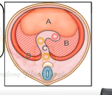
Ans. B - B
Q4. Structure passing through the marker B

- Inferior vena cava
- Oesophagus
- Aorta
- Morgagni hernia
Ans. Morgagni hernia.
Also Read: Hematopoiesis: The Journey Of Blood Cell Production
Sign up to our PrepLadder app today to learn more about this. Access Video Lectures, digital notes, QBank, and Mock Tests for FREE to ace your NEET PG preparation. Elevate your study experience and gear up for success. Start your journey with PrepLadder today!

PrepLadder
Access all the necessary resources you need to succeed in your competitive exam preparation. Stay informed with the latest news and updates on the upcoming exam, enhance your exam preparation, and transform your dreams into a reality!
Navigate Quickly
Components of the Diaphragm
Septum Transversum
Pericardial Peritoneal Membrane
Transverse Section of Diaphragm
Anterior (central tendon of diaphragm)
Dorsal (the pleuroperitoneal membrane)
The dorsal mesentery of the esophagus
Cervical somites