20 High Yield Anatomy Flashcards NEET PG 2026
Dec 16, 2025

Flashcard 1
Difference between Male and Female Gametogenesis

In both sexes, primordial germ cells are present. Primary germ cell is the cell in the beginning to form the gamete. Primary germ cell gives the Oogonium which then gives the Oocyte, Until this, the cell division that occurs is Mitosis
Primary oocyte enters Meiosis I as (2n, 4N). Primary oocyte is the first cell that undergoes Meiosis 1. Secondary oocyte undergoes Meiosis II to form gametes). Gametes are the mature oocytes that are ready for fertilization. Other cells are polar bodies. First polar body and Second polar body.
These polar bodies undergo apoptosis and degenerate. The ratio in a female is 1:1 (1 primary oocyte → 1 gamete). The ratio in a male is 1:4 (1 primary spermatocyte gives 4 sperms). The process of meiosis starts in utero in a female. The primordial germ cells remain dormant until puberty. Till puberty, meiosis does not happen in a male. Spermatogenesis begins only after puberty. Secondary oocytes complete Meiosis II only if it is fertilized by a sperm.
Flashcard 2
Derivatives of Mesoderm

Mesoderm is divided into:
- o Central / Axial mesoderm
- o Para-axial mesoderm
- o Intermediate mesoderm
- o Lateral plate mesoderm
Axial mesoderm forms the notochord. Later, the notochord contributes to the Nucleus Pulposus of intervertebral disc. Para-axial mesoderm contributes to form the somite. A somite further divides into 3 components:
- Sclerotome- will form the vertebrae and ribs
- Myotome - will form the skeletal muscles of the body.
- Dermatome - will form the dermis in the skin.
Flashcard 3
Types of Epithelia in Head & Neck
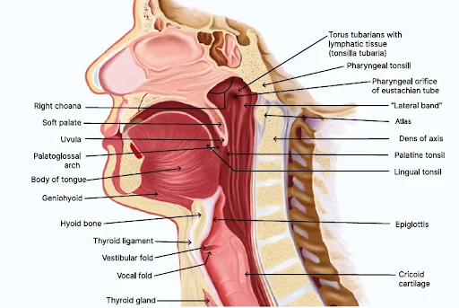
Any external opening in our body is lined by stratified squamous epithelium, like the oral opening, nasal opening, first layer of cornea, external ear canal, etc.
Flashcard 4
Meissner's Corpuscle vs Merkel Cell

Meissner's corpuscle (E) Merkel cell (B) It is an encapsulated receptor, with multiple stacks of cells, for 2-point discrimination. It is a slowly adapting receptor, it detects light touch sensations. It is a rapidly adapting receptor It is a slowly adapting receptor It is seen at the dermo-epidermal junction, in the papillary layer of dermis. It is seen in stratum basale. .jpg)
Flashcard 5
Skeletal vs Cardiac vs Smooth Muscles

|
Skeletal muscle |
Cardiac muscle |
Smooth muscle |
|
Under the control of somatic nervous system |
Under the control of autonomic nervous system |
Under the control of autonomic nervous system |
|
Voluntary muscles |
Involuntary muscles |
Involuntary muscles |
|
Striations are quite prominent due to regular arrangement of sarcomeres. |
Less prominent striations due to less regular arrangement of sarcomeres. |
Sarcomeres are not at all regularly arranged, no striations are seen |
quite long.
|
because the gap junctions in the intercalated discs act as the metabolic tunnels and also as electrical synapses allowing the cells to exchange ions and metabolites.
|
prominent gap junctions and desmosomes. |
|
|
Flashcard 6
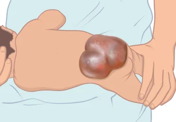
Spina Bifida
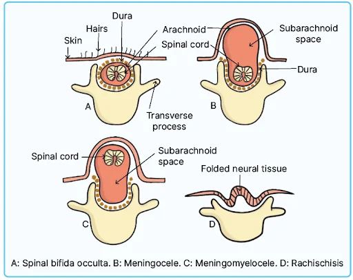
Types of Spina bifida
|
Spina bifida occulta |
|
|
Spina bifida with Rachischisis |
|
|
Spina bifida cystica with Meningocele |
|
|
Spina bifida cystica with Meningomyelocele |
|
- Spina bifida cystica is also known as spina bifida manifesta.
- If the cyst is clear → Meningocele
- If the cyst is not clear and has blue/black markings → Meningomyelocele (neural tissue present).
Flashcard 7
Arterial Supply of the Nervous System

- The circle of Willis is formed at the base of the brain.
- Circle of Willis is contributed by 2 arteries
- Anteriorly: Internal carotid artery (ICA)
- Posteriorly: Vertebral artery (forming the basilar artery)
- Branches of the Internal carotid artery (ICA)
- Anterior cerebral artery: Major artery supplying the medial cerebrum.
- Middle cerebral artery: Supplies the lateral cerebrum.
- Posterior communicating artery
Flashcard 8
Cerebral Arteries

Territory of the middle cerebral artery (mostly lateral cerebrum)
- Broca's motor speech area
- Wernicke's sensory speech area
- Sensory-motor homunculus for the head & the upper limb.
Territory of the anterior cerebral artery
- Mainly medial cerebrum
- Sensory-motor homunculus of the pelvis, perineum & lower limb.
Territory of the posterior cerebral artery
- Occipital visual cortex
- Some part of the parietal sensory cortex.
Flashcard 9
Derivatives of the Pharyngeal Arches


Flashcard 10
Maxillary nerve (C.N. V2)
- It goes towards the maxilla.
- It passes through the foramen rotundum and enters the pterygopalatine fossa.
- Here, the maxillary nerve is related to the pterygopalatine ganglion.
- Pterygopalatine ganglion is topographically related to the maxillary nerve and functionally related to the facial nerve.
- Pterygopalatine ganglion sends post-ganglionic fibres to the lacrimal, nasal & palatine glands.
- Branches of maxillary nerve:
- i. Posterior superior alveolar nerve → Supplies the upper molars
- ii. Infra-orbital nerve
- Infra-orbital nerve is the continuation of the maxillary nerve as it passes through the inferior orbital fissure, in the floor of the orbit.
- The upper teeth & the maxillary sinuses are supplied by the inferior orbital nerve.
- Premolars and anterior upper teeth supplied by anterior superior and middle superior alveolar branches of the infra-orbital nerve.
Flashcard 11
Carotid Sheath
- Contents of the carotid sheath
- Internal carotid artery (ICA) in the upper part; Common carotid artery in the lower part
- Internal jugular vein (IJV)
- Vagus nerve (in the lower part runs in between the ICA & IJV, in the upper part runs behind the ICA & IJV)
- C.N. IX, X, XI in the upper part, later they come out of the sheath and go to their respective places.
- Anterior to the carotid sheath is the ansa cervicalis. It supplies the anterior neck muscles.
- Behind the carotid sheath is the sympathetic trunk, which has stellate ganglion.
Flashcard 12
Superior view of the transverse section of the neck
- Here, we can also see that the pre-tracheal fascia has 2 layers
- Pre-tracheal fascia muscular layer: To cover the anterior neck muscles.
- Pre-tracheal fascia visceral layer: To cover the viscera.
- The visceral layer of the pre-tracheal fascia covers the trachea, oesophagus and the thyroid gland.
- Behind the pharynx and oesophagus, the pre-tracheal fascia modifies into the buccopharyngeal fascia.
- The space behind the buccopharyngeal space is called the retropharyngeal space.
- The carotid sheath is contributed by all the 3 layers of the deep cervical fascia.

Flashcard 13
Oculomotor nerve (C.N. III)

- C.N. III nuclei are present at the level of superior colliculus in the midbrain.
- Edinger-Westphal nucleus
- It is the parasympathetic component
- It controls 2 smooth muscles of the eyeball:
- Sphincter pupillae muscle (for light reflex)
- Ciliaris muscle (for accommodation reflex)
- · Oculomotor nucleus
- It is the motor nucleus (somatic component).
- It controls the skeletal muscles of the eyeball, except lateral rectus muscle (C.N. VI) and the superior oblique muscle (C.N. IV).
- Medial rectus
- Inferior rectus
- Inferior oblique
- Levator palpebrae superioris (LPS)
- Superior rectus
- Lesion in C.N. III
- Ipsilateral down & out eye- because the lateral rectus and the superior rectus muscles are unopposed
- Ipsilateral ptosis- LPS muscle is compromised.
- Ipsilateral fixed dilated pupil (mydriasis)- because of loss of light & accommodation reflex. The sphincter pupillae muscle and the ciliaris muscle are compromised, and the dilator pupillae muscle (supplied by T1 sympathetic fibres) is unopposed.
Flashcard 14
Vertebrae
Human body has 33 vertebrae in total.

Vertebrae
- Cervical - 7
- Thoracic - 12
- Lumbar - 5
- Sacral - 5
- Coccygeal - 4 (Depends on the length)
- Total - 33 vertebra
- Lower thoracic region has more flexion
- Rotation is restricted in the lumbar region because of strong interlocking of the superior and inferior articular facets. They do not provide room for rotatory movement in the lumbar region
- Similarities between Cervical and Lumbar vertebrae that are different from the thoracic vertebrae.
Flashcard 15
Development of embryonic veins

|
Embryonic veins |
Description |
|
Umbilical veins |
|
|
Vitelline veins |
|
|
Cardinal veins |
|
Flashcard 16
Post Development: Adult Positioning of Heart

- Anterior surface of the heart- Sternocostal surface
- Mainly right ventricle
- Partly left ventricle and right atrium
- Left auricle of the left atrium.
- The left atrium relates to the thoracic vertebra and esophagus as it is more posterior.
- The left atrium and descending thoracic aorta can be visualized through transesophageal echocardiography.
- In left atrial hypertrophy, there will be compression of the esophagus, causing dysphagia.
- The right atrium is partly anterior (right ribs) and partly posterior.
- The right border of the heart - Right atrium mainly
- The inferior border of the heart – Right ventricle mainly
- The left border of the heart – Left ventricle mainly (left ribs)
- The superior border of the heart – Left atrium mainly
- The cardiac apex belongs to the left ventricle.
- The left ventricular wall is 3 times thicker than the right ventricular wall. (Ratio- 3:1)
Flashcard 17
Components of TOF
- Pulmonary stenosis
- Overriding of aorta
- Ventricular septal defect
- Hypertrophy of right ventricle
Therefore, TOF + Atrial septal defect: Pentalogy of fallot

Flashcard 18
Phrenic nerve
- Comes from cervical plexus
- Root value → C3 , C4 , C5
- Chief root value → C 4


- Phrenic nerve runs anterior to the scalenus anterior muscle; continues into the thorax region & runs anterior to the hilum of the lung and on lateral side of pericardium to finally reach the diaphragm.
- Phrenic nerve is the only motor supply to diaphragm
- Phrenic nerve is sensory to
- Mediastinal pleura
- Pericardium
- Central portion of diaphragm
- Central portion of peritoneum under central diaphragm
- Therefore, nerve can carry pain of pericarditis, mediastinal pleuritis & central peritonitis.
Flashcard 19
Schematic Diagram of the Brachial Plexus

- Roots join to form the trunks
- Upper trunk: C5 , C6 (supplies the muscles of shoulder, scapula & arm)
- If compromised → Erbs palsy
- Middle trunk: C7
- Lower trunk: C8 , T1 (supplies the distal muscles like hand muscles)
- If compromised → Klumpke's paralysis i.e., the lumbricals & interossei muscles are compromised.
- Upper trunk: C5 , C6 (supplies the muscles of shoulder, scapula & arm)
Flashcard 20
Serratus Anterior Muscle

- Origin: Lateral surface of the upper 8 ribs
- Insertion: Medial border of scapula anteriorly
- Action
- Protraction of scapula
- Overhead abduction of the arm (works along with trapezius muscle) the shoulder joint
Download the PrepLadder App and get the best NEET PG online coaching with world-class video lectures also in हिंglish, QBank, Mock Tests and more!
Download PrepLadder's best app for neet pg preparation for Android
Download PrepLadder's best app for neet pg preparation for ios

PrepLadder Medical
Get access to all the essential resources required to ace your medical exam Preparation. Stay updated with the latest news and developments in the medical exam, improve your Medical Exam preparation, and turn your dreams into a reality!
Navigate Quickly
Flashcard 1
Difference between Male and Female Gametogenesis
Flashcard 2
Derivatives of Mesoderm
Flashcard 3
Types of Epithelia in Head & Neck
Flashcard 4
Meissner's Corpuscle vs Merkel Cell
Flashcard 5
Skeletal vs Cardiac vs Smooth Muscles
Flashcard 6
Spina Bifida
Types of Spina bifida
Flashcard 7
Arterial Supply of the Nervous System
Flashcard 8
Cerebral Arteries
Flashcard 9
Derivatives of the Pharyngeal Arches
Flashcard 10
Maxillary nerve (C.N. V2)
Flashcard 11
Carotid Sheath
Flashcard 12
Superior view of the transverse section of the neck
Flashcard 13
Oculomotor nerve (C.N. III)
Flashcard 14
Vertebrae
Flashcard 15
Development of embryonic veins
Flashcard 16
Post Development: Adult Positioning of Heart
Flashcard 17
Components of TOF
Flashcard 18
Phrenic nerve
Flashcard 19
Schematic Diagram of the Brachial Plexus
Flashcard 20
Serratus Anterior Muscle
PrepLadder Version X for NEET PG
Avail 24-Hr Free Trial