Placenta Formation (Extra Embryonic Mesoderm, Yolk Sac)
Feb 27, 2023

Placenta formation is an important topic in anatomy, as it is a complex process that involves the development and function of the placenta during pregnancy. The placenta is a vital organ that connects the developing fetus to the mother's uterine wall, allowing for the exchange of nutrients, oxygen, and waste products. The placenta also plays a crucial role in hormone production, immune protection, and fetal development.
In the NEET PG Exam, questions related to placenta formation may be asked to test the student's understanding of the anatomy and physiology of the placenta, as well as its role in fetal development and pregnancy. A thorough understanding of placenta formation is essential for medical students.
Read this blog further to get a quick overview of this important anatomy topic for NEET PG exam preparation.
Previous Year Question
Question: FALSE STATEMENT regarding placenta & membranes?
- Tertiary villi seen on day 10
- Fetal circulation established at Day 17-21
- Placenta develops from decidua basalis
- Placenta develops from chorion frondosum
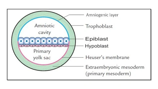
- EMBRYOBLAST CONSISTS OF
- HYPOBLAST → dorsal, degenerate & contributes to extra embryonic structures
- EPIBLAST → ventral columnar cells
- DORSAL AMNIOTIC CAVITY
- VENTRAL YOLK SAC CAVITY
Previous Year Question
Question: Origin of extra embryonic mesoderm?
Answer: Cells lining the primary yolk sac> hypoblast / trophoblast /epiblast

Extra Embryonic Mesoderm
- EXTRA EMBRYONIC MESODERM [EEM] splits to form EXTRA EMBRYONIC coelomic CAVITY which is connected by CONNECTING STALK [connecting stalk forms PRIMARY UMBILICAL CORD]
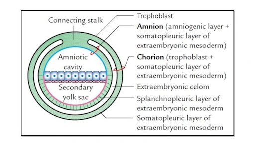
Amnion
It covers amniotic cavity and is Contributed by Amniogenic cells [from trophoblast] and Somatopleuric layer of EEM.
Chorion
It is CONTRIBUTED BY cytotrophoblast that is Syncytiotrophoblast and somatopleuric layer of EEM

Extra Embryonic Coelomic Cavity
It divides the extra embryonic mesoderm into two parts. One towards the yolk sac is called as visceral or splanchnopleuric extra – embryonic mesoderm and beyond that lines the amniotic cavity and outside is called the parietal or somatopleuric extraembryonic mesoderm (towards body wall). Between somatopleuric extraembryonic and visceral/ splanchnic mesoderm is the extra embryonic coelomic cavity. Chorion formed by trophoblast with a somatopleuric layer of extraembryonic mesoderm. Amnion formed by amniogenic cells lining the amniotic cavity and somatopleuric layer of extraembryonic mesoderm. Connecting stalks later become umbilical cord components.
Yolk Sac
A membranous sac linked to an embryo called the yolk sac is made up of cells from the hypoblast layer of the bilaminar embryonic disc. The yolk sac is a far more common name for this, however the Terminologia Embryologica (TE) also calls it the umbilical vesicle. The yolk sac plays a crucial role in the early blood supply of human embryos, and during the fourth week of embryonic development, a large portion of it is integrated into the primordial gut.
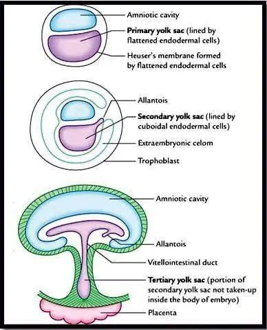
- 1° YOLK SAC → lined by flattened Endodermal cells
- 2° YOLK SAC → lined by cuboidal Endodermal cells
- 3°YOLK SAC → formed during cephalo caudal development of Embryo →forms gut tube → Part of the yolk sac remaining outside the Embryo - 3 YOLK SAC
- In the beginning, we had a primary yolk sac. Later it will become secondary yolk sac and then tertiary yolk sac. Tertiary yolk sac communicates with midgut as vitello – intestinal duct.
- Extra – Embryonic coelomic cavity forming around the baby, there is cephalo- caudal folding of baby. Head comes towards the tail and in this process an amniotic cavity surrounds the body of the baby all around.
- Allantois [Hindgut diverticulum] and the Vitello intestinal duct [midgut diverticulum] will enter the umbilical cord to become its contents.
- PLACENTAL COMPONENTS
- Placenta formed by fetal & maternal contributions
- Placenta formation: Decidua basalis from the maternal side and from the fetal side is chorion frondosum. Chorion layer develops some villi called chorionic villi and they will penetrate into decidua basalis of the maternal side.
- Maternal placenta is decidua basalis [endometrium of uterus] and chorionic villi from fetal placenta component.
UTERUS
- MATERNAL PLACENTA
- DECIDUA BASALIS [DB] → The endometrium where the embryo implants
→forms the maternal /uterine placenta - DECIDUA CAPSULARIS → Surrounds the embryo on luminal side
DONOT FORM PLACENTA - DECIDUA PARIETALIS →The rest of the gravid endometrium
- FETAL PLACENTA
- Derived from chorion
- CHORION FRONDOSUM - chorion towards DB forms layer like projections into it
- CHORION LAEVE - chorion on the side of D. capsularis, DO NOT FORM PLACENTA
- Uterine cavity = Space b/w decidua basalis & Parietalis
- Chorionic cavity = Chorion & Amnion
Chorionic Villi
The chorionic villi are small finger-like extensions of placental tissue that have the same genetic makeup as the developing foetus. Depending on the family history and the availability of lab testing at the time of the surgery, testing may be available for further genetic abnormalities and illnesses.

| PRIMARY VILLUS [Day 12] | Core of cytotrophoblast cells, covered by syncytio TB |
| SECONDARY VILLUS [Day 13-15] | Cytotrophoblast layer invaded by extra Embryonic mesoderm |
| TERTIARY VILLUS [Day 17-21] | Fetal blood vessels invades the mesoderm |
Learn more about this important anatomy topic with the help of our detailed video lectures on the PrepLadder app. You will also get access to comprehensive study notes and high-yield MCQs for practice.

PrepLadder Medical
Get access to all the essential resources required to ace your medical exam Preparation. Stay updated with the latest news and developments in the medical exam, improve your Medical Exam preparation, and turn your dreams into a reality!
Navigate Quickly
Extra Embryonic Mesoderm
Amnion
Chorion
Extra Embryonic Coelomic Cavity
Yolk Sac
UTERUS
Chorionic Villi
Top searching words
The most popular search terms used by aspirants
- NEET PG Anatomy
PrepLadder Version X for NEET PG
Avail 24-Hr Free Trial