Rapid Revision Reignite Anatomy: Question-Answer Format
Sep 23, 2025

Types of Joints and Epiphysis
Big Question 1: What are the types of joints in the human body?
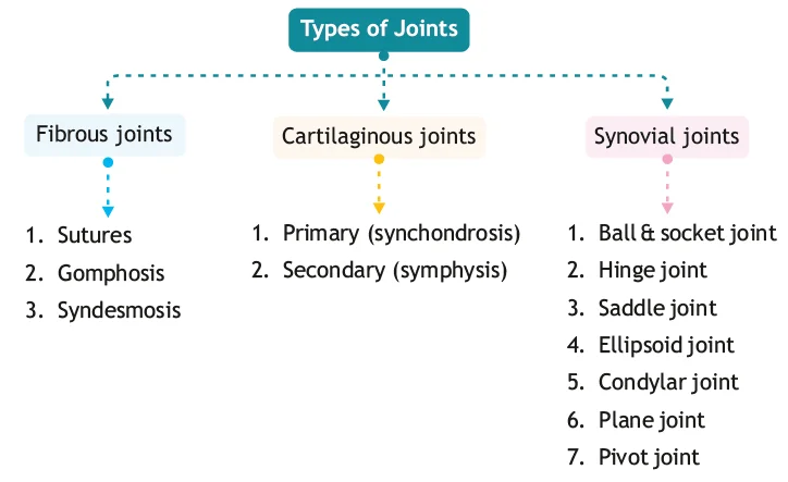
Broad Answer: Joints are classified structurally into fibrous, cartilaginous, and synovial types. Each category has specific subtypes based on the type of tissue and movement permitted.
Detailed Questions
Q1.1: How are joints broadly classified?
Answer: Joints are classified based on the type of tissue uniting the articulating bones into:
- Fibrous joints
- Cartilaginous joints
- Synovial joints
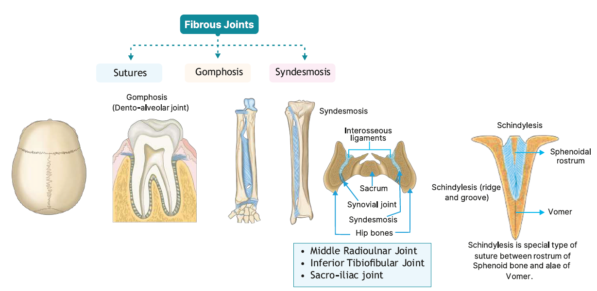
Q1.2: What are the types and subtypes of fibrous joints?
Answer:
| Type of Fibrous Joint | Description | Examples |
| Sutures | Found only in the skull. Bones are united by short collagen fibres immovable joint. | Coronal, sagittal, lambdoid |
| Syndesmosis | Bones connected by a sheet of fibrous tissue. Joint is with the help of interosseous membrane. | Inferior tibiofibular joint, Schindylesis Middle radio-ulnar joint, Sacroiliac joint. |
| Gomphosis | Peg-in-socket joint. | Between tooth and alveolus |
Q1.3: What are the types and subtypes of cartilaginous joints?
Answer:
| Type of Cartilaginous Joint | Other Name | Characteristics | Examples |
| Primary cartilaginous joint | Synchondrosis | United by hyaline cartilage, temporary (fuses later) | Between epiphysis and diaphysis of long bone Costochondral joint: Between riband costal cartilage. Spheno-occipital joint. |
| Secondary cartilaginous joint | Symphysis | United by fibrocartilage, permanent, slight movement | Intervertebral disc, pubic symphysis, manubriosternal joint Xiphisternal joint, Xiphisternal joint |
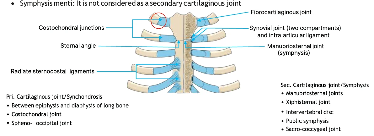
All symphyses occur in the midline (mandibular, manubriosternal, pubic and intervertebral) and all except the mandibular symphysis occur in the postcranial skeleton and resist synostosis. The mandibular symphysis (symphysis menti) is histologically different from the other symphyses; however, the widespread use of this descriptive term ensures that it remains, perhaps inappropriately, within this category.
Big Question 2: What is an epiphysis and what are its types?
Broad Answer: An epiphysis is the end part of a long bone, initially growing separately from the shaft and later fusing through a secondary ossification centre. It contributes to the articulation and growth of the bone.
Detailed Questions
Q2.1: What is an epiphysis?
Answer:
- It is the end part of a long bone.
- It develops from a secondary centre of ossification.
- It is responsible for articulation and growth in bone length.
Q2.2: What are the types of epiphysis?
Answer:
| Type | Description | Example(s) |
| Pressure epiphysis | Takes part in joints and transmits weight | 1. Head of humerus 2. Head of femur 3. Condyles |
| Traction epiphysis | Does not form part of joint; site of muscle attachment Formed due to the pull of the muscle | Trochanters of femur, tubercles of humerus, epicondyles |
| Atavistic epiphysis | Bone that was once separate in ancestors, now fused to main bone | Coracoid process of scapula Os trigonum |
| Aberrant epiphysis | Epiphysis that is not always present; often avariant | Head of first metacarpal, base of remaining metacarpals |
Neuroanatomy
Big Question 3: What are the anatomical features, extent, coverings, and modifications of the spinal cord and cerebrospinal fluid system?
Broad Answer:
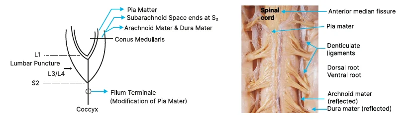
- The spinal cord extends from the foramen magnum to the lower lumbar region and is surrounded by three meninges.
- It contains two enlargements and terminates at L1 in adults.
- The pia mater forms special extensions like the filum terminale and ligamentum denticulatum.
- CSF is present in the subarachnoid space, ideal for lumbar puncture, and flows through the ventricles and central canal of the spinal cord.
Detailed Questions
Q3.1: What is the extent of the spinal cord in adults and infants?
Answer:
- Adult: C1 to lower border of L1
- Infant: C1 to upper border of L3
Q3.2: What are spinal cord enlargements and their levels?
Answer:
- Cervical enlargement: C4 to T2
- Lumbar enlargement: L2 to S3
- Note: Extents are in terms of spinal segments
Q3.3: What is the termination point of the spinal cord and subarachnoid space in adults?
Answer:
- Spinal cord: Middle third of the body of L1 vertebra
- Subarachnoid space: Ends at S2
Q3.4: What is the significance of the subarachnoid space?
Answer:
- Located between arachnoid and pia mater
- Contains CSF
- Ideal site for lumbar puncture (done between L3 and L4)
Q3.5: What are the modifications of the pia mater in the spinal cord?
Answer:
| Structure | Description |
| Filum Terminale | Fine thread-like extension from conus medullaris to coccyx |
| Linea Splendens | Pia mater extension into the anterior median fissure |
| Ligamentum Denticulatum | Tooth-like extensions of pia on either side (2 total), 21 projections each side |
Big Question 4: What are the structural features of brain ventricles and cerebrospinal fluid flow, and what is their clinical relevance?
Broad Answer:
- The brain has four ventricles (two lateral, third, and fourth), which produce and circulate CSF.
- CSF flows from lateral ventricles - foramen of Monro - 3rd ventricle - aqueduct of Sylvius 4th ventricle central canal and subarachnoid space.
- Any blockage (e.g., aqueductal stenosis, Dandy-Walker syndrome) leads to hydrocephalus.
- Each ventricle has specific horns and anatomical landmarks, including the choroid plexus, fornix, thalamus, and hippocampus.
Detailed Questions
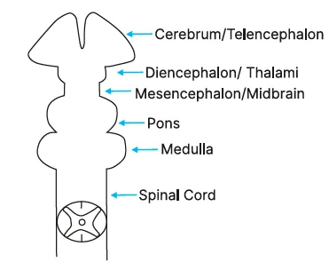
Q4.1: What are the ventricles of the brain and their embryological locations?
Answer:
● Lateral ventricles - in telencephalon
● 3rd ventricle - in diencephalon
● 4th ventricle - in rhombencephalon
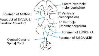
Q4.2: How does CSF flow through the ventricles?
Answer:
Lateral ventricle
↓
Foramen of Monro
↓
3rd ventricle
↓
Aqueduct of Sylvius
↓
4th ventricle
↓
Foramina of Luschka (lateral) and Magendie (medial)
↓
Central canal of spinal cord and subarachnoid space
Q4.3: Where is CSF formed and drained?
Answer:
- Formation: Choroid plexus in lateral ventricles
- Drainage:
- Via arachnoid granulations
- Into dural venous sinuses
- Mainly into superior sagittal sinus
Q4.4: What are the horns and important anatomical relations of the lateral ventricle?
Answer:
| Horn | Anatomical Features |
| Anterior | In frontal lobe |
| Superior | In parietal lobe |
| Posterior | Roof & lateral wall: Tapetum fibres Bulb of posterior horn : Forceps major Calcar avis: Calcarine sulcus |
| Inferior | Roof: Tail of caudate nucleus; Floor: Hippocampus |
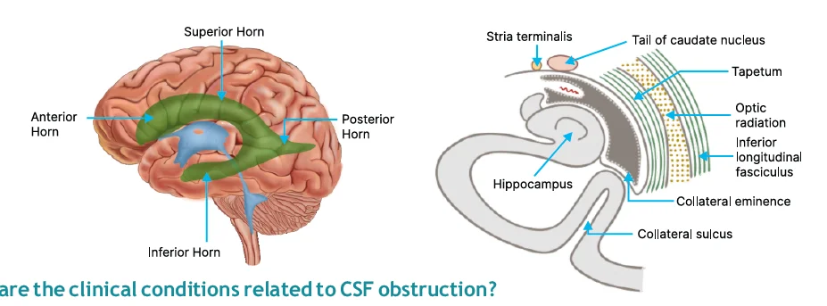
Q4.5: What are the clinical conditions related to CSF obstruction?
Answer:
| Condition | Cause |
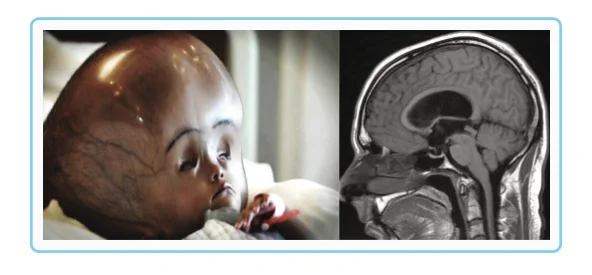
| Aqueductal stenosis | Blockage of aqueduct of Sylvius (cerebral aqueduct) - dilated 3rd ventricle Most common cause of congenital hydrocephalus: Congenital Aqueductal stenosis |
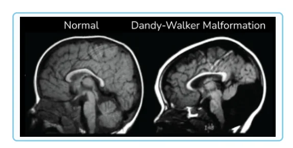
| Dandy-Walker syndrome | Atresia of Luschka and Magendie → dilated 4th ventricle |
| Hydrocephalus | Increased CSF pressure due to obstruction or impaired drainage |
Aqueductal stenosis
Dandy-Walker syndrome
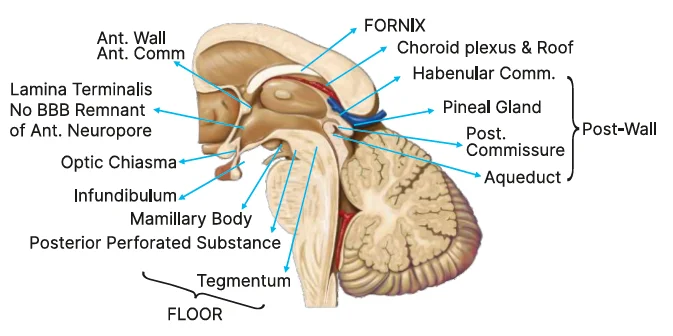
Q4.6: What are the anatomical boundaries and walls of the 3rd ventricle?
Answer:
|
Wall |
Structures |
|
Anterior wall |
|
|
Posterior wall |
|
|
Floor |
|
|
Lateral wall |
|
.jpg)
Thorax
Big Question 5: Discuss the thoracic duct in detail: its course, anatomical relations, termination, and the areas not drained by it.
Broad Answer: The thoracic duct is an important lymphatic channel that plays a crucial role in draining lymph from large parts of the body back into the bloodstream. It begins deep in the abdomen, travels upward through the thorax, and ends by emptying into major veins near the neck. While it drains most regions, there are a few parts of the body that it does not drain.
Detailed Questions
Q5.1: What is the course of the thoracic duct?
Answer: Largest lymphatic vessel
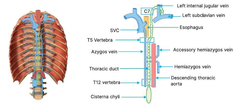
Begins from the cisterna chyli located at L2
↓
Crosses the diaphragm through the aortic opening at the level of T12
↓
Deviates towards the left at the level of T5
↓
Drains lymph at the junction of the left subclavian vein and the left internal jugular vein at the level of C7
Q5.2: What are the areas not drained by the thoracic duct?
Answer:
- The right head and neck
- Right thoracic wall
- Right upper limb
- Right lung
- The right surface of the heart
- Convex part of the liver
If you’re looking to strengthen your final prep, don’t miss out on Rapid Revision Reignite in Question-Answer format by PrepLadder. It’s designed to help Medical PG aspirants cover the entire syllabus quickly with concise notes in a Question-Answer format, high-yield MCQs, and expert-led revision videos—perfect for last-minute reinforcement before the exam.
Download the PrepLadder app now and unlock a 24-hour FREE trial of premium high-yield content. Access Smarter Video Lectures also in हिंglish, Game Changing Qbank, Audio QBank, Structured Notes, Treasures, Mock test for FREE to ace your NEET PG preparation. Elevate your study experience and gear up for success. Start your journey with PrepLadder today!

PrepLadder Medical
Get access to all the essential resources required to ace your medical exam Preparation. Stay updated with the latest news and developments in the medical exam, improve your Medical Exam preparation, and turn your dreams into a reality!
Navigate Quickly
Types of Joints and Epiphysis
Detailed Questions
Neuroanatomy
Detailed Questions
Thorax
Detailed Questions
Top searching words
The most popular search terms used by aspirants
- NEET PG Anatomy
- NEET PG Anatomy Preparation
- NEET PG Preparation
- Rapid Revision