Blistering disorders of the Skin - NEET PG Dermatology
Apr 10, 2023

Blistering disorders are a group of skin conditions characterized by the formation of blisters, which can be a presenting feature of a wide range of dermatological diseases. These conditions include autoimmune blistering diseases, infectious disorders, drug reactions, and inherited disorders. Understanding the clinical presentation, diagnostic criteria, and management of blistering disorders is essential for dermatologists to diagnose and manage these conditions accurately.
Let’s learn more about this important dermatology topic for NEET PG exam preparation.

Blisters
- Fluid filled lesions
- < 0.5 cm called vesicles and > 0.5 cm is called bullae.
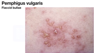
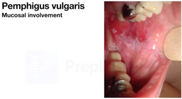
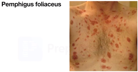
Pemphigus
- Autoimmune disorder
- IgG antibodies are directed against Desmoglein (Dsg) causing intraepidermal blister.
- Erosion with crusting may be seen.
| Pemphigus foliaceus | Pemphigus vulgaris (m/c) | |
| Defect | Dsg 1 (mainly in skin) | Dsg 3 (mainly in buccal mucosa) but late in disease Dsg 1 can also get involved |
| Separation at level of | St. Granulosum | St. spinosum |
| Bullae | Superficial (subcorneal) | Deep (suprabasal) |
| Easily ruptures | Yes | No |
| Involvement | Skin | Initially buccal mucosa and later skin |
| Sites | Seborrheic areas (scalp, retroauricular areas, nasolabial folds, forehead, nose, Presternal, Inter scapular area, upper trunk area | Buccal mucosa, scalp, trunk & extremities |
Clinical signs
- Nikolsky sign - Tangential pressure on bulla causes epidermal separation.
- Bullae spread sign/ Asboe Hansen sign - Perpendicular pressure in bulla causes it to spread / increase in size.
- They are positive in both but PV > PF.
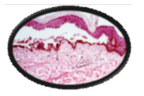
Histology
- Acanthocytes
- Row of tombstones appearance

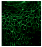
Direct Immunofluorescence (DIF)
- Fish net like patterns due to intercellular deposition of Anti DSG Ab.
Treatment
- Steroids
- Cyclophosphamide
- Rituximab (Anti- CD20)
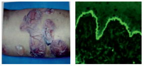
Bullous Pemphigoid
- lgG against BP Ag2 > BP Ag1.
- C/F - Tense sub epidermal bullae thus do not rupture easily and the fluid gets absorbed so appears as settled bullae.
- Histology - Sub epidermal split with eosinophilic collection.
- Direct Immunofluorescence (DIF) - Linear IgG & complement deposits in Basement membrane Zone.
| Pneumonic | PEMPHIGUS | PEMPHIGOID |
| M - Mucosal involvement I - Intraepidermal blister N - Nikolsky sign A - Acanthocytes R - Row of tombstone app | + | - |
Direct Immunofluorescence (DIF)
Pemphigus
Pemphigoid
Linear IgA Disease
- IgA against BP Ag 2
- Bimodal presentation
- Child (< 5 years) - Face (perioral), groin (perianal).
- Adults (60 years) - Trunk & extremities.
- Grouped blisters around erythematous plaques.
- String of pearl /bead or cluster of jewel appearance.
- DIF - Linear IgA deposits in BMZ (Basement Membrane Zone).
- DOC - Dapsone
Chronic Bullous Disease Childhood
- Childhood variant of linear IgA disease
Dermatitis Herpetiformis
- Gluten hypersensitivity
- Associations
- Celiac disease
- HLA B8, DQ2, DR3
- Anti -Endomysial Antibodies
- Anti- Transglutaminase - 3 Antibody
- C/F - Extremely pruritic vesico papules, later on erosions seen over extensors
- Histology - Papillary tip microabscesses
- DIF - Granular IgA deposits in dermal papilla
- DOC - Dapsone
- TOC - Gluten free diet
| Avoid | Can take |
| B - Barley | Rice |
| R - Rye | Maize |
| O - Oat (least amount of gluten) | Ragi |
| W - Wheat |
Herpes Gestationis
- Autoimmune
- C/F - Periumbilical lesions develops in females in 3rd Trimester
- Self-limiting condition
- Fetal mortality is high - 30%
- DIF - Linear C3> IgG deposits in Basement Membrane Zone
- DOC - Steroid

Epidermolysis Bullosa
- Also known as mechanobullous disorder.
- Occurs at the site of trauma, friction & pressure handling.
| Types | |
| Congenita | Acquisita |
| Congenital | Acquired |
| Early | Late |
| Common | Rare |
| Genetic | Immunobullous |
Subtypes of Epidermolysis bullosa congenita
- EB Simplex - defected K5/14
- EB Junctional - defected Laminin
- EB Dystrophicans - defected Collagen 7
Dermatology Related Articles:
Dermatology Related Articles:
Darier’s Disease And Hailey-Hailey’s Disease
Feature Darier’s Disease Hailey – Hailey’s Disease Inheritance Autosomal Dominant Autosomal Dominant Defect Ca2+ ATPase Ca2+ ATPase Gene ATP 2A2 ATP 2C1 Site Seborrheic areas Intertriginous areas (skin folds) like axilla, groin & inframammary folds Lesion Verrucous (warty lesion), greasy with sand paper feel Recurrent, flaccid, vesicular (may rupture causing erosions) Histology Acantholysis, dyskeratosis with corps ronds, corps grains Acantholysis, dyskeratosis with dilapidated brick wall appearance


Erythema Multiforme And Epidermal Necrosis
Feature Erythema Multiforme Epidermal Necrosis Cause Viral infection > Drugs Drugs Lesion Target lesions/Iris lesion/ Bull’s eye lesions Targetoid lesions
Epidermal separation as per body surface area
<10% - Steven Johnson syndrome
10 – 30% (overlap syndrome)
> 30% - TENNikolsky sign Absent Present Mucosal involvement Absent present


Hemorrhagic crusting (SJS)
Epidermal separation (TEN)
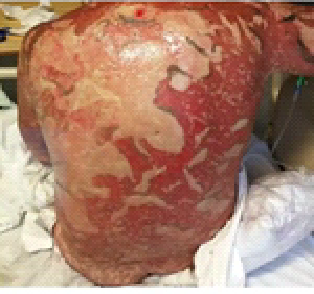
TEN
- Toxic Epidermal Necrolysis
- Sheet like epidermal separation
- Poor prognosis
Previous Year Questions
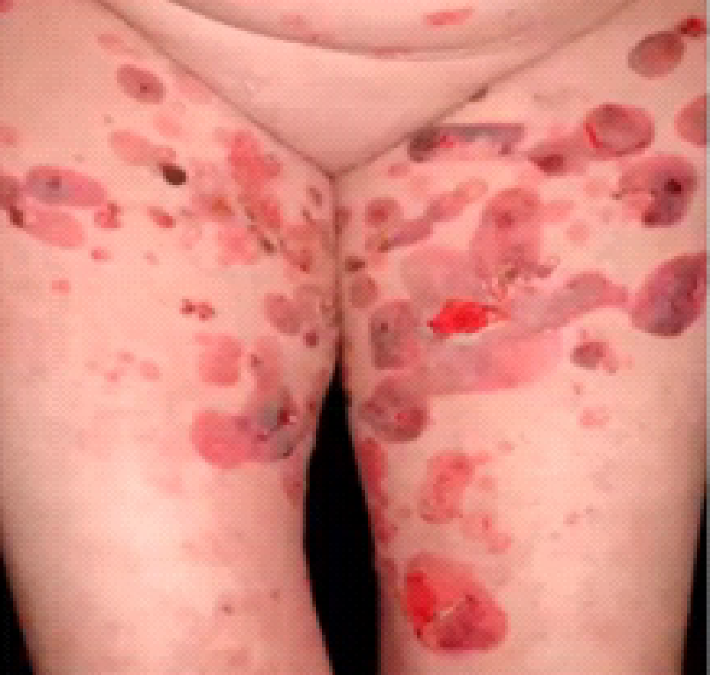
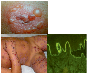
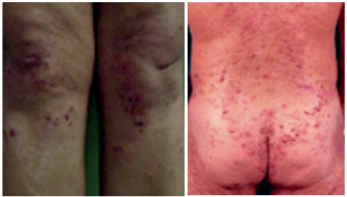
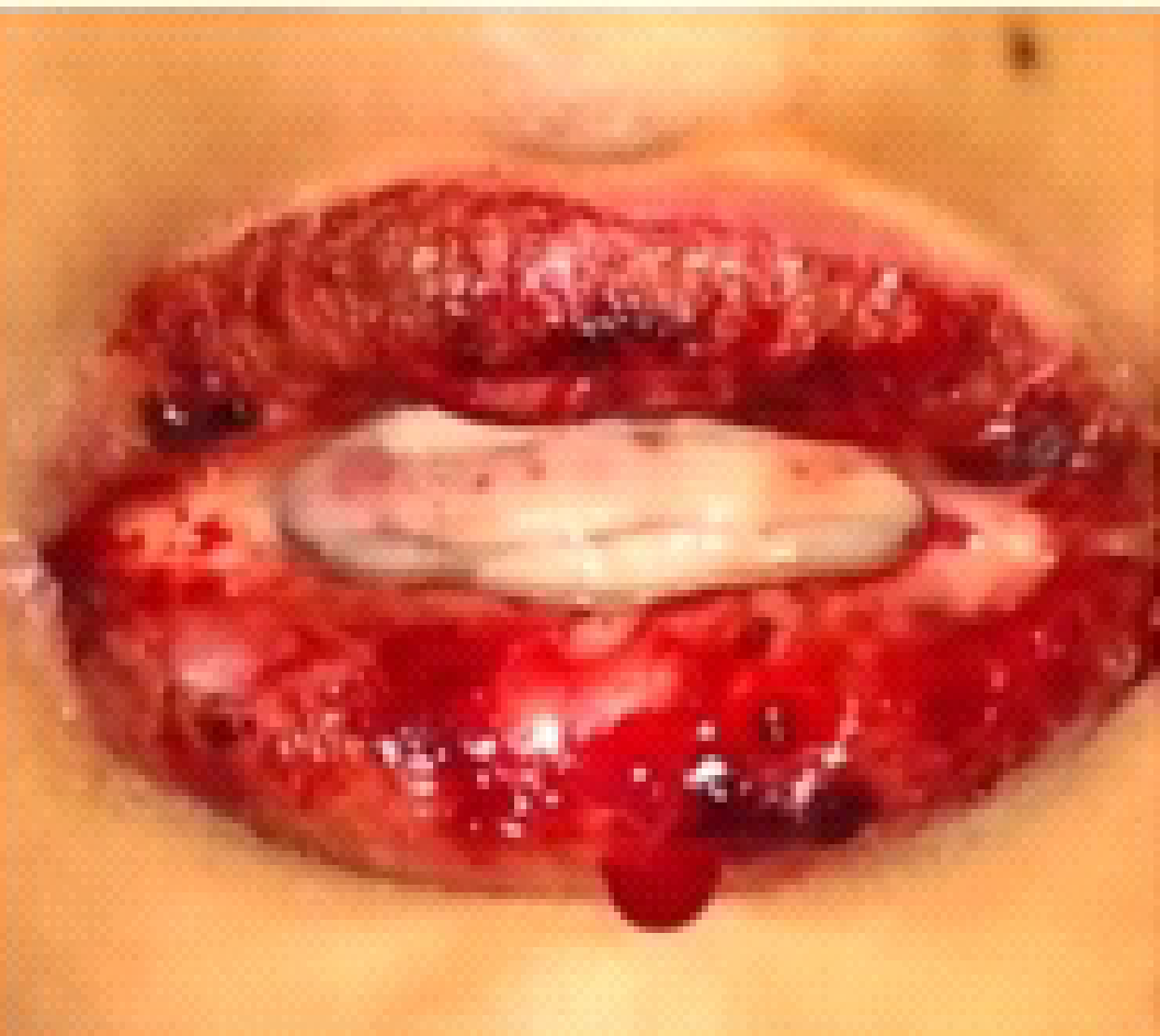
Q. Patient presents with oral lesions along with flaccid blisters on scalp, face, trunk and extremities, as shown in the image. Diagnosis? (AIIMS NOV 2019)
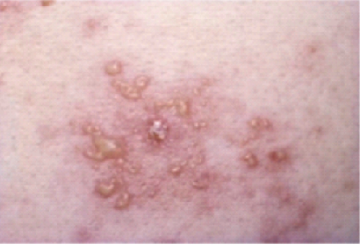
- Pemphigus vulgaris
- Pemphigus foliaceous
- Dermatitis herpetiformis
- Bullous pemphigoid
Q. Sub epidermal blisters with neutrophils, treatment of choice? (INICET Nov 2020)
- Rituximab
- Dapsone
- Cyclosporine
- Azathioprine
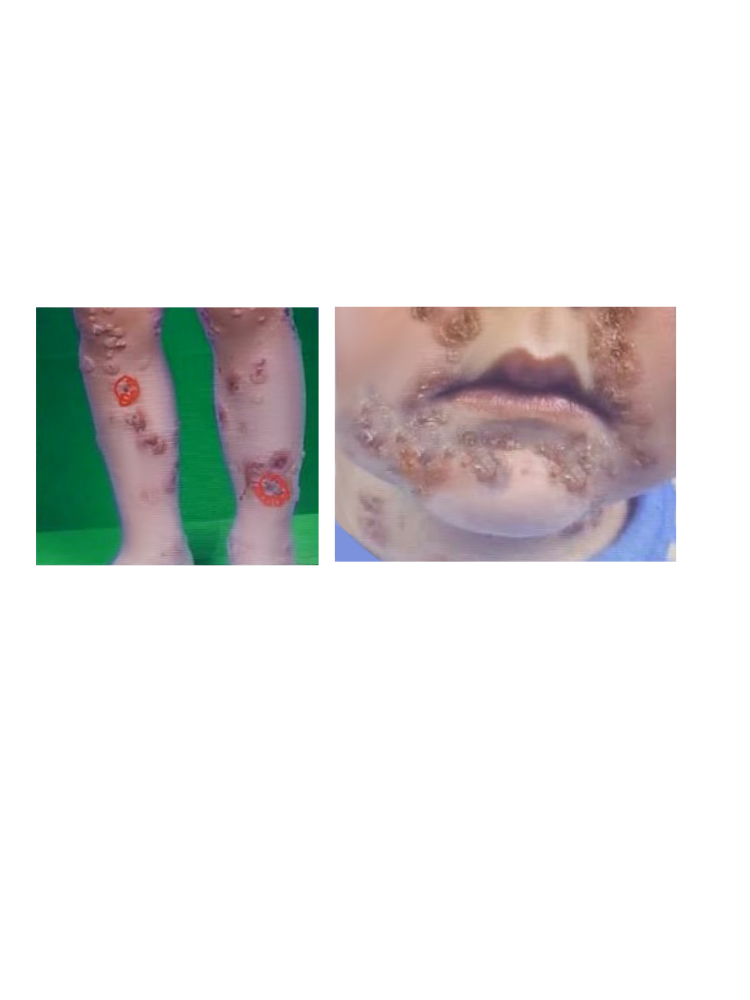
Q. A 10-year-old child comes with erosions and blisters on trauma prone areas. There is a history of death of a sibling after 10 days with similar illness. The most probable diagnosis is? (AIIMS JUNE 2020)
- Epidermolysis bullosa
- Pemphigus
- Congenital syphilis
- Chronic bullous disease of childhood
Q. Erythema multiforme is seen in? (NEET PG 2019)
- Celiac disease
- ITP
- Antimalarials
- Leukemia
Q. Arrange superficial to deep blister disease? (AIIMS MAY 2019)
- Pemphigus vulgaris
- Linear IgA
- Bullous pemphigoid
- Epi. Dystrophica
Ans - A>C>B>D
Q. A patient with known case of T.B has been taking multiple medications and now he has presented with the given image, what is the most appropriate diagnosis? (FMGE dec 2020)
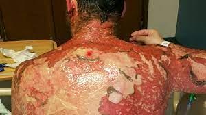
- Fixed drug eruption
- TEN
- SJS
- Scrofuloderma
To study this high-yield dermatology topic in detail, download the PrepLadder app and watch engaging video lectures by our expert faculty.

PrepLadder Medical
Get access to all the essential resources required to ace your medical exam Preparation. Stay updated with the latest news and developments in the medical exam, improve your Medical Exam preparation, and turn your dreams into a reality!
Navigate Quickly
Blisters
Pemphigus
Clinical signs
Histology
Direct Immunofluorescence (DIF)
Treatment
Bullous Pemphigoid
Direct Immunofluorescence (DIF)
Pemphigus
Pemphigoid
Linear IgA Disease
Chronic Bullous Disease Childhood
Dermatitis Herpetiformis
Herpes Gestationis
Epidermolysis Bullosa
Subtypes of Epidermolysis bullosa congenita
Darier’s Disease And Hailey-Hailey’s Disease
Erythema Multiforme And Epidermal Necrosis
Hemorrhagic crusting (SJS)
Epidermal separation (TEN)
TEN
Previous Year Questions
Q. Patient presents with oral lesions along with flaccid blisters on scalp, face, trunk and extremities, as shown in the image. Diagnosis? (AIIMS NOV 2019)
Q. Sub epidermal blisters with neutrophils, treatment of choice? (INICET Nov 2020)
Q. A 10-year-old child comes with erosions and blisters on trauma prone areas. There is a history of death of a sibling after 10 days with similar illness. The most probable diagnosis is? (AIIMS JUNE 2020)
Q. Erythema multiforme is seen in? (NEET PG 2019)
Q. Arrange superficial to deep blister disease? (AIIMS MAY 2019)
Q. A patient with known case of T.B has been taking multiple medications and now he has presented with the given image, what is the most appropriate diagnosis? (FMGE dec 2020)