INI-CET Image-Based Questions: High-Yield Images & Topics to Revise
Sep 8, 2026
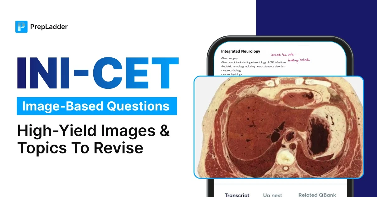
INI-CET is not an exam where knowing the textbook alone is enough. A question can present the same concept through a chest X-ray, CT image, histopathology slide, ECG, clinical photograph, gross specimen, instrument, or anatomical diagram and expect you to recognise the key visual clue before arriving at the answer.
The supplied references consistently describe image-based questions as a major part of INI-CET preparation and emphasise that image interpretation is a trainable skill rather than something that depends on natural visual memory.
The important point is that you do not need to memorise an endless collection of individual pictures. You need to build a high-yield visual vocabulary: recognise the classic pattern, know its closest differential, and connect the image to the underlying concept.
This guide brings those image-heavy areas together subject-wise so that your revision is focused on the pictures most worth knowing before the exam.

Why Image-Based Questions Matter in INI-CET
INI-CET covers a wide visual range. Radiology, pathology, ECGs, clinical photographs, ophthalmology images, surgical instruments, gross specimens, microbiology microscopy, and anatomical images can all form the basis of questions.
The question may also go beyond simple identification. After showing an image, the examiner can ask for the most likely diagnosis, the next investigation, definitive treatment, causative organism, inheritance pattern, or another associated finding.
This is why simply looking at images repeatedly is not enough. The better approach is to look at an image and actively ask yourself what you are seeing, what it represents, what can mimic it, and what the examiner could ask next.
The Eight Visual Categories to Cover
Before going subject by subject, keep these major image families in your revision plan:
| Image category | Core areas to revise |
| Radiology | X-rays, CT, MRI, ultrasound and classic radiological signs |
| Pathology | Histopathology, cytology and gross specimens |
| Clinical photographs | Dermatology, syndromic facies and bedside clinical signs |
| Ophthalmology | Fundus, anterior segment and external eye photographs |
| ECG | Rhythm, axis, conduction abnormalities and ischaemic patterns |
| Instruments | Surgical, obstetric, gynaecological and anaesthesia instruments |
| Microbiology | Microscopy, special stains and culture morphology |
| Anatomy | Cadaveric images, diagrams and radiological anatomy |
These categories cover the main recurring visual formats described across the reference material.

Download INI CET Previous Year Question Papers PDF For Free
Radiology: Start With the Highest-Yield Visual Patterns
Radiology deserves early attention because it has a broad image vocabulary spanning plain films, CT, MRI, and ultrasound. The key is to learn recognisable signs and patterns, rather than attempting to memorise every possible image.
High-Yield Radiology Revision List
Imaging area High-yield images and patterns Chest X-ray Pulmonary oedema, hyperinflation, Kerley B lines Cardiac imaging Classic cardiac silhouettes Mediastinum Anterior, middle and posterior compartment masses HRCT chest Tree-in-bud and crazy-paving patterns CT brain Hyperdense and hypodense intracranial lesions Abdominal CT Appendicitis, intussusception and major vascular emergencies Ultrasound Gallstones, renal pathology and important obstetric landmarks Urological imaging Stones, obstruction and characteristic IVU/urogram appearances
The supplied material specifically highlights pulmonary oedema versus hyperinflation, mediastinal compartments, Kerley B lines, tree-in-bud, crazy paving, CT brain density patterns and classic abdominal CT diagnoses.
A useful way to revise radiology is to train yourself to identify the dominant sign first. For example, do not immediately jump to a disease name on seeing a chest X-ray. First identify the key radiographic pattern, then map that pattern to the most likely diagnosis.

Pathology: Learn the Signature Finding
Pathology is another major image-heavy area. Questions may show a microscopic field and expect you to identify a characteristic cell, inclusion, arrangement, or tissue pattern. Gross specimens can also be tested through their colour, cut surface, or characteristic morphology.
High-yield pathology images
Signature image Classic association Reed-Sternberg cell Hodgkin lymphoma Psammoma bodies Papillary thyroid carcinoma and other classic associations Aschoff body Rheumatic carditis Russell bodies Plasma cells Keratin pearls Squamous cell carcinoma Signet-ring cells Gastric carcinoma Caseating granuloma Tuberculosis Mallory-Denk bodies Alcohol-associated liver disease Bread-and-butter appearance Fibrinous pericarditis Nutmeg liver Chronic venous congestion
These signature associations are specifically represented in the reference material.
The revision strategy here should be image → hallmark structure → diagnosis → associated fact. Do not stop after naming the slide.
For example, if you recognise a characteristic inclusion or cell, immediately revise the disease association, common differential, and one important clinical correlation that can be tested in the same question.
Medicine and ECG: Combine Visual Recognition With Clinical Reasoning
Medicine image questions are particularly useful because the image is often only the first step. A smear, endoscopic image, pathology image, or ECG may be followed by a question about diagnosis, complication, or management.
The supplied medicine examples include schistocytes on peripheral smear, Mallory-Denk bodies, normal sinus rhythm, Crohn disease endoscopy, and characteristic clinical findings.
High-Yield ECG patterns
ECG What to recognise Additional revision Atrial fibrillation Irregularly irregular rhythm, absent P waves Rate/rhythm management Complete heart block AV dissociation Immediate management WPW Short PR and delta wave Associated arrhythmias and treatment STEMI Territory-specific ST elevation Culprit artery LBBB Characteristic QRS morphology Clinical significance
The reference specifically identifies STEMI territory mapping, complete heart block, atrial fibrillation, WPW, and LBBB as priority ECG patterns.
That means ECG review should not end at “identify the rhythm.” For every classic tracing, know what you do next.
Clinical Photographs and Dermatology: Train Your Eye for Morphology
Dermatology is particularly suited to image-based preparation because the appearance itself often carries the diagnosis. Reading a description of a rash is not equivalent to seeing it repeatedly.
High-yield clinical photographs
Image finding Diagnosis/association Heliotrope rash Dermatomyositis Gottron papules Dermatomyositis Malar rash sparing nasolabial folds SLE Target lesions Erythema multiforme Nikolsky sign Pemphigus vulgaris Salmon-pink rash Still disease Annular plaque with central clearing Dermatophytosis
These examples are specifically identified as high-yield dermatological image patterns in the supplied reference.
When revising a dermatology image, avoid focusing only on the disease name. Train yourself to identify:
Morphology → distribution → colour → surface → characteristic sign → diagnosis
That method is much more useful when the exam presents an unfamiliar-looking but conceptually related photograph.
Ophthalmology: Fundus Images Need Pattern Recognition
Ophthalmology image questions commonly depend on recognising the anatomical structure first and then identifying the abnormality.
Fundus and eye images to revise
Image Key association Papilloedema Raised intracranial pressure Disc pallor Optic atrophy Diabetic retinopathy Characteristic retinal vascular changes Hypertensive retinopathy Retinal vascular changes and advanced signs Retinal detachment Detached retinal appearance Corneal ulcer Characteristic corneal morphology Anterior-segment photographs Corneal, angle and lid pathology
The supplied sources specifically include disc pallor, papilloedema, diabetic and hypertensive retinopathy, retinal detachment, and anterior-segment findings among recurring ophthalmology images.
The key is to identify where the abnormality is located before deciding what it represents.
Surgery: Revise the Image, Sign and Instrument Together
Surgery image-based questions often combine an imaging finding with a clinical presentation and then ask for the diagnosis, investigation, or definitive management.
The supplied surgery reference demonstrates this pattern through renal stones, urographic appearances, ectopic ureter, catheter identification, cystography, and liver CT findings.
High-yield Surgery images
Area Image or finding to revise Common question angle Urolithiasis Staghorn calculus Stone composition Renal/urinary TB Calcification, strictures and destructive changes Diagnosis/complications Renal pelvis lesions Classic urographic signs Diagnosis/investigation Congenital urology Duplex system, ectopic ureter Diagnosis Catheters Catheter appearance Indication and use Trauma CT cystogram patterns Site/type of bladder injury Liver lesions Triphasic CT appearances HCC vs metastasis and enhancement pattern Surgical instruments Retractors, clamps and scopes Identification and procedure
For example, the reference uses a branched renal calculus with recurrent UTI and alkaline urine to test recognition of a struvite stone, while an IV urographic appearance is used to test a congenital urinary abnormality.
A useful surgical revision rule is:
Image finding → anatomical location → diagnosis → definitive management
Do not memorise the picture in isolation. Know what the image means clinically.
Pediatrics: Images Can Test Development as Well as Disease
Pediatrics has an important difference from adult clinical subjects: the image may be a developmental milestone, growth curve, dysmorphic feature, fundus photograph, or neonatal radiograph, rather than a conventional diagnostic scan.
The supplied pediatric material includes ventral suspension as a milestone clue, growth-pattern graphs, Tay-Sachs disease, craniosynostosis, and Apert syndrome.
High-Yield Pediatrics Images
Image type What to revise Developmental photographs Milestones and examination manoeuvres Growth curves Somatic, brain, lymphoid and gonadal growth patterns Fundus photographs Classic metabolic/storage disease findings Skull photographs Craniosynostosis patterns Dysmorphic photographs Apert and other syndromic appearances Neonatal X-rays Congenital diaphragmatic hernia and other emergencies Skin photographs Common neonatal and pediatric rashes Fontanelle diagrams Anterior and posterior fontanelle landmarks
One example from the source is particularly useful for revision: the ventral suspension image is linked with the milestone of neck holding at around 3 months.
Another common exam style is matching a physical appearance to a syndrome. The supplied example uses craniosynostosis with a characteristic “mitten hand” deformity to identify Apert syndrome.
This means pediatric image preparation should cover both “What is this?” and “At what age/pattern does this occur?”
Microbiology: Revise the Organism by Its Visual Signature
Microbiology image questions are usually built around recognisable morphology rather than complicated images. The key categories include stained smears, microscopy, culture plates, and special preparations.
High-yield Microbiology images
Image Visual clue to recognise Gram stain Gram reaction + bacterial morphology Ziehl-Neelsen stain Acid-fast bacilli KOH preparation Fungal elements India ink Cryptococcus Culture plate Characteristic colony morphology Wet mount Motility/organism morphology Parasite images Eggs, cysts or trophozoites
The important strategy is to connect appearance with organism and test. For every image, revise the associated specimen, stain, organism, and one key clinical association.
Anatomy: The Image May Ask for a Structure Rather Than a Diagnosis
Anatomy image questions are often deceptively simple. A labelled structure, cadaveric photograph, or cross-sectional image may require precise three-dimensional orientation.
High-yield Anatomy image areas
Area Revision focus Gross anatomy Identification of important structures Neuroanatomy Brainstem, cranial nerves and cross-sections Embryology Developmental anomalies represented visually Radiological anatomy CT/MRI anatomical landmarks Cadaveric images Relations and surface anatomy Applied anatomy Structures relevant to clinical procedures
The supplied references recommend focusing on clinically relevant anatomical landmarks rather than trying to memorise every named structure.
A useful approach is to practise anatomy images without labels first. Identify the structure independently, then check the answer.
Obstetrics and Gynaecology: Ultrasound, Instruments and Classic Clinical Images
For OBGYN, image preparation should cover three broad areas: ultrasound, clinical photographs, and instruments.
High-yield OBGYN image revision
Category What to revise Obstetric ultrasound Fetal and placental landmarks Early pregnancy imaging Ectopic pregnancy and related findings Pelvic imaging Adnexal and uterine pathology Gynaecological specimens Classic gross appearances Instruments Obstetric and gynaecological instruments Clinical photographs Characteristic external findings
The reference material explicitly includes obstetric and gynaecological tools within the expected image categories.
The key is not just identifying an instrument or ultrasound finding. Know its indication, associated condition, and important complication because the image can be only the opening clue to the question.
Orthopaedics: Learn Classic X-Ray Patterns
Orthopaedic image questions are primarily about recognising alignment, fracture configuration, bone changes, and characteristic radiographic patterns.
High-yield Orthopaedics images
Topic Visual revision target Fractures Classic named fracture patterns Dislocations Joint alignment Bone tumours Characteristic radiographic appearance Osteomyelitis Typical bone changes Rickets Metaphyseal changes Joint disorders Recognisable imaging patterns Pediatric orthopaedics Growth plate and developmental abnormalities
The supplied syllabus specifically identifies fractures, pediatric orthopaedics, joint diseases and prosthetics as important orthopaedic areas.
When looking at an orthopaedic image, first establish which bone, which region, and which alignment is abnormal before trying to recall a named diagnosis.
ENT: Focus on What the Examiner Can Show You
ENT image revision should concentrate on photographs and visual examinations rather than attempting to turn every topic into an image question.
Revise:
- Otoscopic images
- Tympanic membrane abnormalities
- Nasal and sinus imaging
- Laryngeal photographs
- Oral and pharyngeal lesions
- Head and neck masses
- Classic clinical signs
The broader image-category reference specifically includes clinical photographs and oral/ENT findings among the visual formats that can appear.
Forensic Medicine and Anaesthesia: Small Sections, High Recall Value
These subjects should be kept concise but should not be omitted.
Forensic Medicine
Focus on:
- Injury patterns
- Burns
- Ligature marks
- Toxicology-related images
- Weapon/instrument identification
- Autopsy findings
The supplied syllabus places medico-legal procedures, toxicology and autopsy findings within Forensic Medicine.
Anaesthesia
Revise:
- Airway devices
- Endotracheal tubes
- Laryngoscopy equipment
- Breathing systems
- Perioperative monitors
- Characteristic monitoring tracings
The reference set specifically includes anaesthesia devices within the instrument/equipment image category.
The Same Image Can Generate More Than One Question
This is an important distinction for INI-CET preparation.
Do not revise an image as:
Image = diagnosis
Instead, revise it as:
Image → finding → diagnosis → differential → association → next step
For example, a renal stone image may lead to a question on its composition; an ECG may lead to management; a pathology slide may lead to the associated disease; an instrument may lead to its indication.
The reference material specifically recommends learning patterns with their differentials and linking the visual finding to the underlying concept.
INI-CET Image-Based Questions: High-Yield Images & Topics to Revise
The Five-Step Method for Solving an Unknown Image
When an image looks unfamiliar in the exam, do not immediately search your memory for a disease name. A fixed reading sequence reduces anchoring and helps you extract the clue that actually matters.
1. Identify the modality
First determine what you are looking at:
X-ray, CT, MRI, ultrasound, ECG, histopathology slide, gross specimen, clinical photograph, instrument or anatomical diagram.
Also check orientation, side markers, ECG labels, staining method, and relevant technical details when available.
2. Scan the complete image
Do not focus immediately on the most obvious abnormality.
Use a consistent sweep appropriate to the image. In a chest X-ray, for example, assess the trachea and mediastinum, cardiac silhouette, lung fields, hila, costophrenic angles, and bones/soft tissues. A systematic sweep helps prevent an incidental finding from becoming the diagnosis.
3. Find the dominant visual clue
Ask yourself:
“What is the single most important abnormal feature in this image?”
A classic sign, characteristic cell, specific rash morphology, ECG abnormality, or distinctive anatomical relationship usually provides the shortest route to the answer.
4. Map the finding to the diagnosis
Now connect the visual clue with your stored pattern vocabulary.
Do not stop at recognition. Think about the closest differential and the feature that separates it from the correct answer. The supplied references specifically recommend pattern recognition together with differential diagnosis rather than memorising isolated images.
5. Read exactly what the question asks
The image may make the diagnosis obvious, but the question may ask for something else.
It could ask for:
- The next investigation
- Definitive management
- Causative organism
- Associated condition
- Complication
- Inheritance pattern
- Histological feature
This distinction is explicitly highlighted in the reference material.
Common Image-Based Question Traps
Knowing classic images is only half the job. INI-CET can make a familiar image difficult by placing the useful clue next to a misleading one.
| Common trap | What happens |
| Anchoring | You identify the first obvious abnormality and stop looking |
| Incidental finding | A secondary finding distracts from the main diagnosis |
| Normal variant | A normal appearance is mistaken for pathology |
| Wrong modality or sequence | CT/MRI or imaging sequence is misidentified |
| Stem bias | The clinical history makes you interpret the image prematurely |
| Wrong question | You identify the diagnosis correctly but answer a management question incorrectly |
These are among the common visual errors described in the supplied reference material.
A simple rule helps:
Look at the image independently first. Then use the stem to refine the interpretation.
How to Build Strong Image-Recognition Memory
Randomly scrolling through hundreds of pictures may create familiarity without creating reliable recall. The references favour active testing, repeated exposure, and connecting the picture with the underlying concept.
Use the Cover–Recall–Explain Method
For every image:
Cover the caption → identify the finding → name the diagnosis → explain the hallmark clue → state the likely next question.
For example, after seeing a histopathology slide, do not merely say “Hodgkin lymphoma.” Ask yourself:
What cell am I seeing? What is its defining appearance? What other diagnosis could resemble it? What associated fact could be tested?
The same principle applies to ECGs, radiographs, skin photographs, and instruments.
Build a One-Page Image Notebook
A useful final-revision format is:
| Column | What to record |
| Image | Actual image |
| Hallmark clue | Single defining visual feature |
| Diagnosis | Most likely diagnosis |
| Differential | Closest mimic |
| Exam twist | Investigation, management, association or complication |
This keeps image preparation focused and prevents notes from becoming another textbook.
Revise Images by Pattern, Not by Source
Do not organise your revision only according to where you found an image. Organise it according to visual pattern.
For example:
All chest X-rays together → all fundus photographs together → all ECGs together → all histopathology signatures together → all dermatology photographs together.
The supplied references specifically recommend grouping images by category and using repeated exposure to develop comparative visual memory.
This is especially useful for confusing pairs and differentials.
| Pattern group | Compare |
| Chest X-ray | Pulmonary oedema vs hyperinflation |
| ECG | AF vs other irregular rhythms |
| Histology | Different inclusion bodies |
| Dermatology | Similar-looking rashes |
| CT brain | Hyperdense vs hypodense lesions |
| Urology | Different stone appearances |
| Pediatric skull | Different craniosynostosis patterns |
Final High-Yield Image Checklist
Before the exam, make sure your final revision has covered these major visual buckets:
| Priority | What should be familiar |
| Very high | Chest X-ray patterns |
| Very high | CT brain and classic abdominal CT |
| Very high | Histopathology signature cells and structures |
| Very high | Gross pathology specimens |
| Very high | ECG rhythms, conduction defects and STEMI patterns |
| Very high | Dermatological photographs |
| Very high | Fundus photographs |
| High | Surgical instruments and urological imaging |
| High | Microbiology stains and microscopy |
| High | Pediatric syndromes, milestones and growth curves |
| High | Obstetric ultrasound and gynaecological images |
| High | Orthopaedic X-rays |
| High | Anatomy and radiological anatomy |
| Must not skip | ENT, Forensic Medicine and Anaesthesia visual material |
The sources consistently identify radiology and pathology as the major visual contributors, with ECGs and clinical photographs also important, while instruments, ophthalmology, microbiology and anatomy should not be neglected.
Frequently Asked Questions
How image-heavy is INI-CET?
The supplied references describe INI-CET as having a higher visual load than NEET-PG, but they do not support assigning one fixed number of image-based questions to every session. The exact proportion can vary.
Which subjects should I prioritise for image-based preparation?
Start with Radiology and Pathology, then give strong attention to ECG/Medicine, Dermatology, Ophthalmology and Surgery. Continue with Pediatrics, Microbiology, Anatomy, OBGYN and the other specialties so that blind spots do not remain.
Should I memorise every image I see?
No. The more efficient strategy is to learn the classic pattern vocabulary and understand the feature that distinguishes similar images.
What should I do when I have never seen the image before?
Identify the modality, systematically scan the image, isolate the dominant feature, form the closest diagnostic pattern, and then read what the question actually asks. This approach allows you to reason even when the exact image is unfamiliar.
Is solving image-based MCQs better than simply reading an atlas?
For exam preparation, active image-based practice has an important advantage because it forces you to commit to an interpretation before seeing the answer. The supplied references recommend active recall rather than passive viewing.
Does negative marking change the way I should approach image questions?
It should make you more deliberate, not automatically make you skip visual questions. The supplied reference notes that partial elimination can improve decision-making under the −1/3 marking scheme for MD/MS.
Conclusion
INI-CET image-based questions become much less intimidating once image revision is treated as a separate skill.
The goal is not to remember an enormous atlas. It is to recognise the hallmark visual pattern, distinguish it from its closest mimic and connect it immediately with the underlying clinical concept.
Revise images subject-wise, practise them through active recall, compare confusing patterns side by side, and train yourself to ask one question after every diagnosis:
“What else can the examiner ask from this image?”
That is what turns image recognition from passive familiarity into exam-ready recall.

PrepLadder
Access all the necessary resources you need to succeed in your competitive exam preparation. Stay informed with the latest news and updates on the upcoming exam, enhance your exam preparation, and transform your dreams into a reality!
Navigate Quickly
Why Image-Based Questions Matter in INI-CET
The Eight Visual Categories to Cover
Download INI CET Previous Year Question Papers PDF For Free
Radiology: Start With the Highest-Yield Visual Patterns
High-Yield Radiology Revision List
Pathology: Learn the Signature Finding
High-yield pathology images
Medicine and ECG: Combine Visual Recognition With Clinical Reasoning
High-Yield ECG patterns
Clinical Photographs and Dermatology: Train Your Eye for Morphology
High-yield clinical photographs
Ophthalmology: Fundus Images Need Pattern Recognition
Fundus and eye images to revise
Surgery: Revise the Image, Sign and Instrument Together
High-yield Surgery images
Pediatrics: Images Can Test Development as Well as Disease
High-Yield Pediatrics Images
Microbiology: Revise the Organism by Its Visual Signature
High-yield Microbiology images
Anatomy: The Image May Ask for a Structure Rather Than a Diagnosis
High-yield Anatomy image areas
Obstetrics and Gynaecology: Ultrasound, Instruments and Classic Clinical Images
High-yield OBGYN image revision
Orthopaedics: Learn Classic X-Ray Patterns
High-yield Orthopaedics images
ENT: Focus on What the Examiner Can Show You
Forensic Medicine and Anaesthesia: Small Sections, High Recall Value
Forensic Medicine
Anaesthesia
The Same Image Can Generate More Than One Question
INI-CET Image-Based Questions: High-Yield Images & Topics to Revise
The Five-Step Method for Solving an Unknown Image
4. Map the finding to the diagnosis
5. Read exactly what the question asks
Common Image-Based Question Traps
How to Build Strong Image-Recognition Memory
Use the Cover–Recall–Explain Method
Build a One-Page Image Notebook
Revise Images by Pattern, Not by Source
Final High-Yield Image Checklist
Frequently Asked Questions
How image-heavy is INI-CET?
Which subjects should I prioritise for image-based preparation?
Should I memorise every image I see?
What should I do when I have never seen the image before?
Is solving image-based MCQs better than simply reading an atlas?
Does negative marking change the way I should approach image questions?
Conclusion
Top searching words
The most popular search terms used by aspirants
- INI CET High Yield Questions
- INI CET Image Based Questions
- INICET Preparation Strategy and Tips