Abnormal Pupils - NEET PG Ophthamology
Feb 10, 2023

Abnormal pupillary responses can provide valuable diagnostic information in various medical conditions, including neuro-ophthalmic disorders, systemic diseases and drug effects.
Knowledge of abnormal pupillary responses is crucial for medical professionals appearing for NEET PG as it forms an important part of the ophthalmology section in the exam.
Read this blog and get a quick overview of this important Ophthalmology topic.

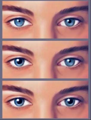
Argyll Robertson Pupil
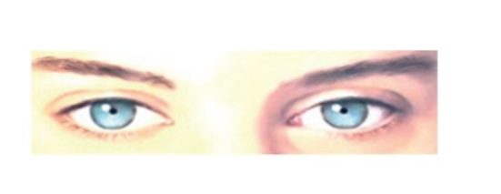
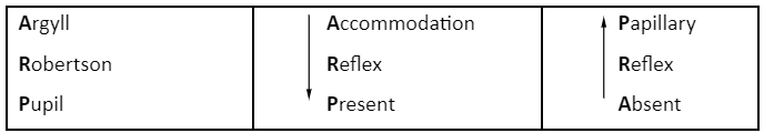
- Argyll Robertson Pupil, also known as light near dissociation.
- Light reflex is absent here, and near reflex is present.
- It is seen in neurosyphilis
- Lesions are seen in internuncial neurons between pretectal and edinger-westphal neurons.· The supranuclear adrenergic fibres being damaged hinders the process of dilatation. It is a continuous parasympathetic stimulation.
- Symptoms
- B/L, constricted, irregular pupils
- Seen in neurosyphilis, tertiary syphilis
- Does not constrict to light but to near vision (because lesion is in dorsal midbrain damaging the light fibers but accommodation center is spared)
- Light reaction - negative; accommodation reflex - positive
- A/k/a Prostitute’s pupil (because it accommodates but no reaction)
ARP
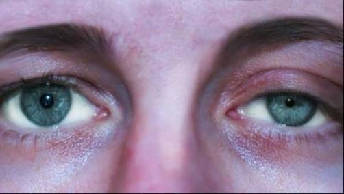
Adie’s Pupil
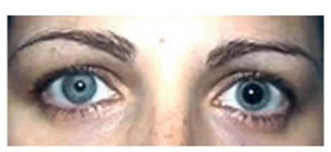
- Seen in young women (80%)
- Seen after viral fever
- U/L dilatation of pupil
- Virus attacks ciliary ganglion, CN III is present in ciliary ganglion which gets affected causing
- Parasympathetic constrictor fibres of pupil to damage, so no constriction
- Unopposed sympathetic dilation of pupil occurs
- Vermiform movement of iris (sluggish moment)
- No reaction to light & Accommodation reflex positive (aberrant regeneration: fibers innervating ciliary body i.e., III nerve fibers and parasympathetic fibers also innervate pupil, but the fibers innervating ciliary body is 30% than the pupil therefore after damage, there is regeneration in ciliary body. Ciliary body fibers goes into aberrant mode & goes to pupil which is why it constricts so strongly to near objects but no reaction to light)
|
Argyll Robertson pupil |
Adie’s pupil |
|
|
- Adie’s Test
- Confirmed by pilocarpine challenge test
- 0.125% pilocarpine solution used, put in both eyes
- Normal pupil does not constrict because solution is very dilute
- Abnormal pupil will constrict
- d/t denervation supersensitivity
- Confirmed by pilocarpine challenge test
Also Read : NEET PG 2023: High-Yield Topics for Ophthalmology
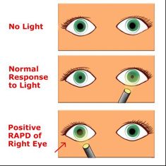
Marcus Gunn Pupil (RAPD)
- Seen in anterior vision pathway (i.e. optic disc, optic nerve just before optic chiasma) disorders like optic neuritis (M/C), AION, optic nerve glioma
- Tested by swinging torch light test
- Take a torch and flash it into normal eye and observe: Pupil constricts in both eyes
- When light is flashed into the affected eye: Pupil dilates in that eye because of nerve fibre damage.

Hutchinson’s Pupil
- Seen in case of post head trauma leading to cerebral compression because of progressive intracranial haemorrhage
- Consists of 3 stages
- Stage 1: Pupil of the side of trauma constricts (due to irritation of oculomotor nerve which carry parasympathetic fibres)→ Normal side unaffected
- Stage 2 (6 hrs. later): Due to progressive intracranial hemorrhage pupil of the injured side become dilated (parasympathetics are lost so unopposed sympathetic dilatation) → Normal pupil constricts because by this time some blood has trickled to other hemisphere and cause irritation of 3rd nerve.
- Stage 3 (after another 6 to 10 hrs): Pupil of both sides fully dilated because parasympathetic fibres of 3rd Nerve on both sides are lost.
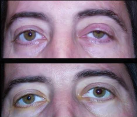
HORNER’S SYNDROME
- Aka Oculosympathetic paralysis
- Triad of:
- Ptosis
- Miosis
- Anhidrosis
- Enophthalmos is:
- Not a true enophthalmos, only apparent / pseudo (no actual sinking of eyeball).
- Because of ptosis (upper lid falls down) and inferior tarsal muscle palsy (inferior lid comes up) causing narrowing of palpebral fissure.
Congenital HS
- Heterochromia: Iris affected is lighter color
- M/C cause: Birth trauma, neuroblastoma
Acquired HS
- M/C cause: Pancoast tumor, carotid dissection (painful)
- Confirmatory Test: Apraclonidine test
- Put drop of Apraclonidine in both eyes and observe the change in Pupil
- HS Pupil dilates, normal doesn’t
Important Information
- Cocaine test is not done now a days used
FAQ’S
1) A case with a fixed, dilated pupil after a head injury, what could be possible diagnosis
A)Oculomotor nerve palsy
2): Anisocoria which is greater in dim light than in bright light
- Adie's tonic pupil
3) In which stage of syphilis MRP may present?
- Teritary syphillis
Did you find this blog insightful? If, yes then download the PrepLadder app and get regular updates on similar medical notes blogs, effective preparation strategies and latest exam updates.

PrepLadder Medical
Get access to all the essential resources required to ace your medical exam Preparation. Stay updated with the latest news and developments in the medical exam, improve your Medical Exam preparation, and turn your dreams into a reality!
Navigate Quickly
Argyll Robertson Pupil
ARP
Adie’s Pupil
Marcus Gunn Pupil (RAPD)
Hutchinson’s Pupil
HORNER’S SYNDROME
Congenital HS
Acquired HS
Top searching words
The most popular search terms used by aspirants
- NEET PG Exam Tips
- NEET PG Ophthamology