Rapid Revision Reignite Orthopedics: Question-Answer Format
Sep 10, 2025
Introduction to Orthopedics Typical & Atypical Fractures
Big Question 1: What is orthopedics? What are the classifications of fractures, and what are the stages of fracture healing?
Broad Answer: Orthopedics is the branch of medicine concerned with the prevention, diagnosis, and treatment of musculoskeletal disorders. Fractures are classified by type, site, and etiology, and they heal through inflammatory, reparative, and remodeling stages.
Detailed Questions
Q1.1: Who coined the term 'orthopedics'?
Answer:
- Orthopedics comes from the Greek words "ortho" (making straight) and "paedics" (child).
- It means "making bent bones of a child."
- The term was coined by Sir Nicholas Andry, who is known as the Father of Orthopedics.
Q1.2: What is a fracture?
Answer: A fracture is a breach in the continuity of a bone, periosteum, or both.
Q1.3: What are the key differences between typical and atypical fractures?
Answer:
|
Typical Fractures |
Atypical fractures |
|
|
Q1.4: What are the stages of fracture union/healing?
Answer:
- The stages are:
- Stage of impaction
- Stage of induction
- Stage of hematoma formation
- Stage of callus formation
- Stage of consolidation
- Remodeling
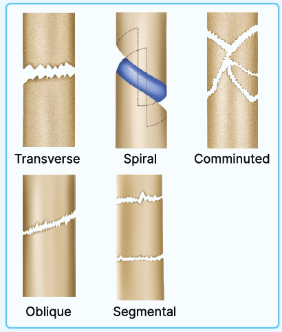
Q1.5: How does the prognosis of a fracture vary with the type of fracture?
Answer:
- The prognosis is best for fractures with the highest raw surface area for healing, which are oblique.
- The prognosis decreases in the following order: oblique (best) > transverse > spiral > comminuted > segmental (worst).
Special X-Ray Views In Ortho
Big Question 2: What are the indications and technique for Coyle's view of the elbow, and how is the scaphoid view performed?
Broad Answer:
- The Coyle's view, also known as the trauma oblique view of the elbow, is performed with the patient seated, elbow flexed at 90°, palm facing down, and the X-ray tube angled 45° lateromedially.
- It is primarily used to detect radial head fractures, elbow dislocations, and to visualize the capitellum of the distal humerus.
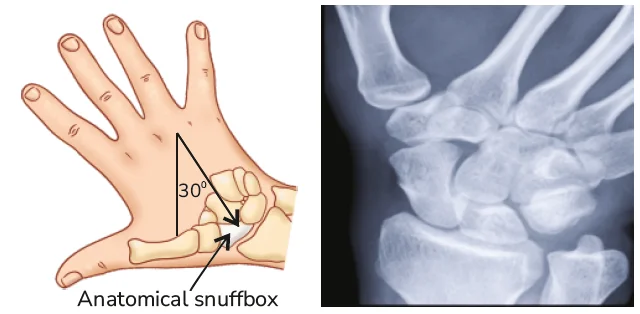
- The scaphoid view involves positioning the wrist in ulnar deviation to eliminate bony overlap and enable clear visualization of the scaphoid bone. This is usually performed as a PA projection, but can also be considered an AP view.
- Both views enhance the detection of subtle fractures and joint abnormalities in the wrist and elbow.
Detailed Questions
Q2.1 What is Coyle's view of the elbow, and what are its clinical indications?
Answer:
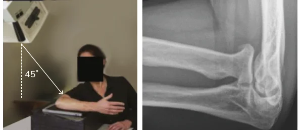
- Also called the Trauma Oblique View of the Elbow
- In the Coyle method, the sitting axial lateromedial projection, where the patient is seated, the elbow is flexed at 90°, palmar side downwards, with the X-ray tube at a 45° angle projection lateromedially.
- The Coyle's view is performed for
- Suspected Radial Head Fracture
- Elbow Dislocation
- Visualise Capitellum of the Distal Humerus
Q2.2 Explain the positioning and purpose of the Scaphoid View in wrist radiography.
Answer:
- The view is performed with the wrist in ulnar deviation to free the scaphoid from bony superimposition.
- Although typically performed as a PA projection, the view may also be described as an AP view.
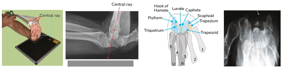
Q2.3 Explain Carpal Tunnel View.
Answer:
- It is used to obtain an axial view of the carpal canal, with particular attention to the hook of the hamate, pisiform bone, and trapezium fractures.
- The hand is maximally dorsiflexed using the opposite hand or a strap.
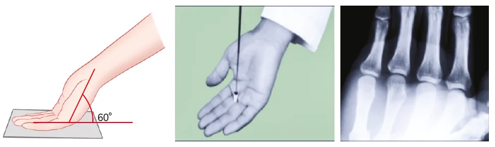
Q2.4 What is Brewerton's View and what is its clinical significance?
Answer:
- The metacarpophalangeal joint is flexed to 65°, with the dorsum of the proximal phalanx flat against the radiograph cassette.
- The beam is angled 15° from ulnar to radial to profile the collateral recesses.
- It is helpful to visualize a collateral ligament Avulsion Fracture.
.jpg)
Malunion & Open Fractures
BIG QUESTION 3: What is malunion of a fracture, what are its types with examples, and what treatment options are available?
Broad Answer: Malunion is the healing of a fracture in an incorrect anatomical position, leading to deformity or functional impairment. It can be classified by angulation, rotation, or shortening, and treatment includes corrective osteotomy, physiotherapy, or supportive measures.
Detailed Questions
Q3.1: What is malunion?
Answer:
- Definition: Fracture fragments unite in an abnormal position.
- Causes: Improper treatment.
- Effects: Shortening, alteration in posture/balance.
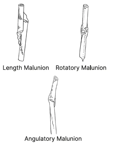
Q3.2: What are the types of Malunion?
Answer:
| Types of Malunion | Description |
| Length Malunion | Other Name: Overlap Malunion Overriding of the proximal and distal fragments So, the length is altered |
| Rotatory Malunion | Proximal and distal fragments are united in the wrong fashion. Normal length is maintained. Alignment of the fragments are altered (Rotated) |
| Angulatory Malunion | Proximal and distal fragment are not present in the straight line (Angulation) |
Q3.3: What are the Treatment options for Malunion?
Answer:
- Malunion of 5° - 10° can be corrected by remodeling, so active treatment is not necessary.
- 3 methods of treatment
- Osteoclasis
- ORIF
- Osteotomy
- Treatment of choice: Corrective osteotomy
| Methods | Explanation |
| Osteoclasis | Voluntary breaking of the bone Bone is broken and the angulation is corrected, and is refixed |
| ORIF (Open reduction internal fixation) | Osteoclasis is done (First) The angular bone is straightened It is fixed with the plate and screws |
| Osteotomy | When Malunion is around the joint. Cutting the wedge of the bone. Correcting the angle and fixing it again. It is known as the corrective osteotomy. It is the gold standard treatment |
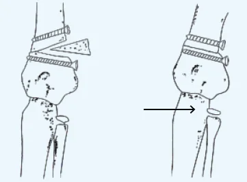
Osteotomy
Bone Tumor
BIG QUESTION 4: What is the classification of bone tumors, and what are the origin, common sites, clinical features, radiological findings, and treatment of osteochondroma?
Broad Answer: Bone tumors are classified as benign or malignant, based on origin, behavior, and histology. Osteochondroma is a common benign bone tumor arising from the metaphysis of long bones, presenting as a painless bony swelling; diagnosis is by X-ray, and treatment is surgical excision if symptomatic.
Detailed Questions
Q4.1: What is the Lichtenstein Classification of bone tumors?
Answer:
|
Cell type |
Benign |
Malignant |
|
Bone |
Osteoid osteoma |
|
|
Cartilage |
|
PYQ: FMGE 2019, 2020 |
|
Fibrous tissue |
Fibroma |
Fibrosarcoma |
|
Bone marrow |
Eosinophilic granuloma (Histiocytosis-X) |
|
|
Vascular |
Haemangioma |
|
|
Uncertain |
Giant cell tumor (osteoclastoma) |
Malignant giant cell tumor |
Q4.2: What are the key features of Osteochondroma?
Answer:
- Also called exostosis.
- Most common primary benign bone tumor.
- It is an offshoot of the spongy bone tissue covered with a cartilaginous cap.
- This is due to the unwanted activity of the cambium layer of the periosteum.
- Most common age :
- Growth period (growth spurt) - teenagers.
- Male preponderance.
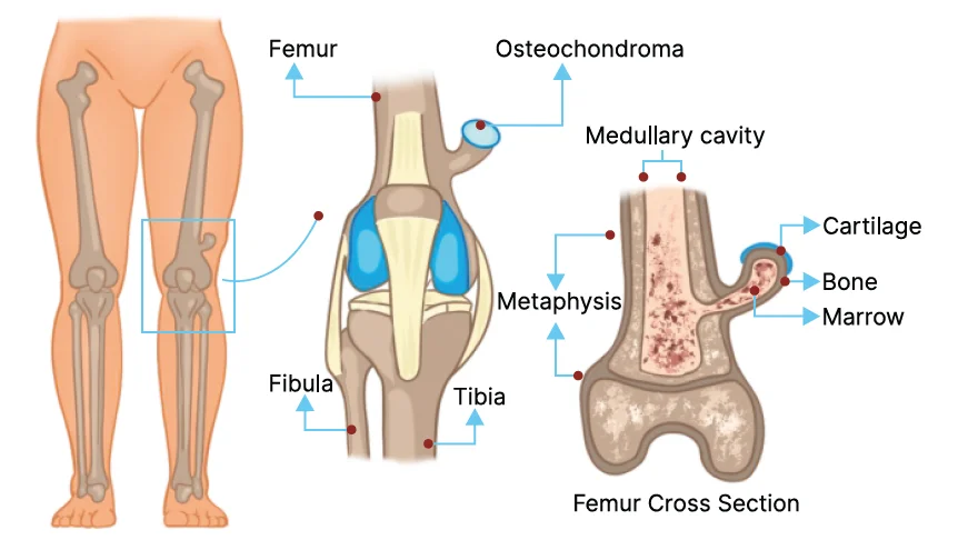
Q4.3: What are the common sites of osteochondroma?
Answer:
Most common site -The lower end of the femur.
2nd common site - Upper end of the tibia. Around the knee joint. Most common around the metaphysis of long bones, the crest of the ilium.
Q4.4: What are the Clinical Presentations of Osteochondroma?
Answer:
- 99% are asymptomatic
- Pain - Pain should not happen in Osteochondroma. If present, the possibilities are:
- Adventitious bursitis
- Fracture of exostosis
- Compression of surrounding nerves - Neuritic pain.
- Malignant change of Osteochondroma - 1% chance.
- Restriction of joint movements
- A firm, tender swelling fixed to the bone
- Gross appearance of exostosis - Cauliflower appearance.
If you’re looking to strengthen your final prep, don’t miss out on Rapid Revision Reignite by PrepLadder. It’s designed to help Medical PG aspirants cover the entire syllabus quickly with concise notes in a Question-Answer format, high-yield MCQs, and expert-led revision videos—perfect for last-minute reinforcement before the exam.
Download the PrepLadder app now to access high-yield content with 24-hr Free Trial. Explore premium study resources like Video Lectures also in हिंglish, digital notes, Audio QBank, and Mock Tests for a seamless exam preparation. Time to begin your NEET PG coaching online with PrepLadder.

PrepLadder Medical
Get access to all the essential resources required to ace your medical exam Preparation. Stay updated with the latest news and developments in the medical exam, improve your Medical Exam preparation, and turn your dreams into a reality!
Navigate Quickly
Introduction to Orthopedics Typical & Atypical Fractures
Detailed Questions
Special X-Ray Views In Ortho
Detailed Questions
Malunion & Open Fractures
Detailed Questions
Bone Tumor
Detailed Questions
Top searching words
The most popular search terms used by aspirants
- NEET PG Orthopedics
- NEET PG Orthopedics Preparation
PrepLadder Version X for NEET PG
Avail 24-Hr Free Trial