Musculoskeletal Radiology: Bone Tumors
Feb 15, 2023

Musculoskeletal Radiology, and in particular, Bone Tumors, is considered an important topic for the NEET PG exam. This is because bone tumors are a common cause of musculoskeletal pain and can impact an individual's mobility and quality of life.
The diagnosis and management of bone tumors often involves the integration of radiology with other specialties such as orthopedics, oncology, and pathology. A good understanding of musculoskeletal radiology, including bone tumors, is essential for effective patient care.
In this blog, you’ll get a brief overview of this important radiology topic for NEET PG exam preparation. Read on.
BONE TUMORS
Epiphyseal Tumors

- Chondroblastoma
- Seen in children (growth plate not fused). Calcification is seen: Chicken wire calcification
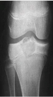
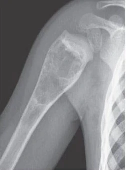
- Giant cell tumor
- Middle aged adult
- Distal end of radius
- Soap bubble appearance
- From epiphysis towards metaphysis
Diaphyseal Tumors
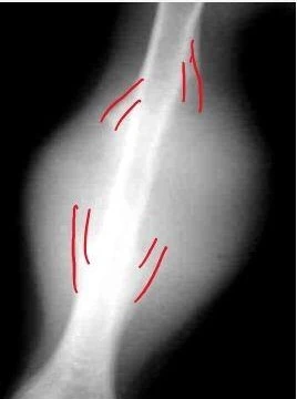
- Adamantinoma
- Chief bone affected : Tibia
- Rare; malignant potential (+)
- Has associated bowing of bone

|
Important Information
|
- Ewing sarcoma
- 0 - 20 yrs [MC in 1st decade]
- Inflammatory (+)
- Onion peel periosteal reaction: Large permeative lesion
|
Important Information
|
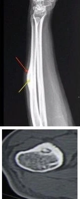
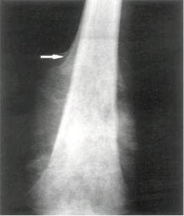
- Osteoid osteoma
- Any age
- Nocturnal pain relieved on NSAIDS [because Nidus releases prostaglandin]
- Diaphyseal tumor in shaft of long bone
- Lesion in cortex; has central lucency k/a Nidus
- Periosteal thickening
- TOC: CT Guided radiofrequency Ablation
Metaphyseal Tumors
- Simple bone cyst
- Proximal humeral end
- Well defined geographical lytic lesion
- Pathological fracture with fragment fallen into the lesion k/a “Fallen Fragment”
- 1st decade
- No septations
- Aneurysmal Bone cyst
- 2nd decade
- Septations (+) (Multiple): Soap bubble appearance
- MRI: Multiple blood – blood / blood – fluid levels (Bleeding inside lesion)
|
Important information Fluid – fluid levels on MRI
|
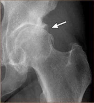
- Osteochondroma / Exostosis
- Bony outgrowth growing away from joint
- Not a true bone tumor
- If multiple lesions are seen – Diaphyseal Aclasis / Hereditary Multiple exostoses
- Risk of developing chondrosarcoma
- IOC : MRI to rule out malignancy [>1.5 cm/2cm]

|
Important Information
|
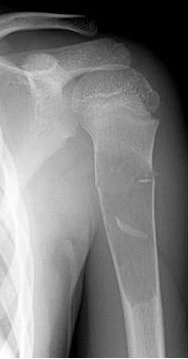
- Osteosarcoma
- 0-20 yrs (2nd decade)
- New bone (+)
- “Codman’s triangle” [lifting up of periosteum]
- “Sunburst” reaction (new bone spicules)
- Aggressive metaphyseal tumor: Has dense matrix (suggestive of new bone formation)
|
Important Information
|
|
Important Information
|
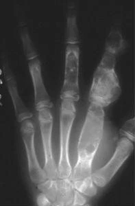
Multiple enchondromas
- Expanded short tubular bones of hand with stippled calcification: Hallmark of enchondroma
- Only multiple enchondroma : OLLIER DISEASE
- Multiple enchondroma + Hemangioma: MAFFUCCI DISEASE
Fibrous dysplasia
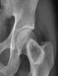

- Ground glass matrix in femoral neck (metaphyseal region)
- If any pathological fracture associated then can see a deformity k/a Shepherd crook deformity (femur is bend)
- Rind sign: Sclerotic rim (femoral neck)
- Fibrous dysplasia can involve multiple bones k/a Multiple / Polyostotic fibrous dysplasia can associated with
- McCune Albright syndrome: Precocious puberty, endocrinopathies
- Mazabraud syndrome: Soft tissue Myxoma
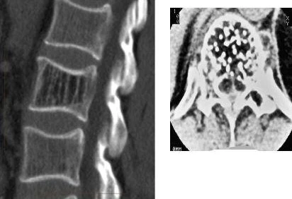
Spinal Hemangioma
- MC benign lesion in vertebral body
- Body trabeculation: “Jail bar / corduroy cloth appearance” [sagittal view]
- Bony trabeculation (cross - section): “Polka dot sign”
Also Read: Why is Radiology the most preferred branch?
ARTHRITIS
Osteoarthritis
- Aka degenerative arthritis
- Large joints affected > Small joints
- Chronic history; Asymmetric (E.g., lateral joint space is more involved)
- No morning stiffness
- Osteophytes (New bone formed)
- Subchondral Sclerosis
Rheumatoid Arthritis
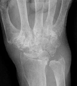
- Small joints > Large joints
- Young female; Morning stiffness (+)
- Symmetrical
- Erosions (marginal): Periarticular Osteopenia
- Sparing : DIP
|
Important Information
|
- Because of constant inflammation synovial proliferation k/a Pannus seen in Rheumatoid arthritis
|
Important Information
|
- Ball Catcher’s view
- Done for RA
- MCP joint are better visualized in this view
- Aka Brewerton’s view

Psoriatic Arthritis

- Gross destruction of bone are seen giving
- Pencil in cup appearance
- Telescoping (+)
- Has associated skin, nail changes [silvery scale, oil pitting nail]
- Very destruction form of psoriatic arthritis k/a Arthritis Mutilans
- Opera glass sign
- MC joint affected : DIP
Gouty Arthritis

- Older Male
- MC joint : 1st MTP
- Large erosions k/a “Rat bite erosion”
- Overhanging margins: “Martel G sign”
- Dual energy CT can identify (Tophi) the presence of uric acid in subcutaneous and in joint space
|
Important Information
|

Neuropathic Joint / Charcot joint

- Joint looks very destructed but patient feels no pain
- MC seen in DM patients
- MC : Foot (mid tarsal joint)
- Other causes
- Leprosy
- Syringomyelia
- Any neuropathy
- X-ray shows 6Ds
- Distention (joint effusion (+))
- Density (increased)
- Debris
- Dislocation
- Disorganization
- Destruction
One liners
- Epiphyseal enlargement seen in
- Juvenile idiopathic arthritis
- Epiphyseal dysgenesis seen in
- Congenital hypothyroidism [Baby will have short stature]
- Widened intercondylar notch seen in
- Hemophilic arthropathy [has squaring of patella]
Also Read: Metabolic Bone Diseases - NEET PG Radiology
Ankylosing Spondylitis
|
Important Information
|

- Young patient (<50 yrs); Pan joint fusion / pan spinal fusion
- Inflammatory arthritis affecting spine
- Most sensitive investigation / IOC for sacroiliitis: MRI [STIR sequence / fat suppressed sequence]
- 1st manifestation: Sacroiliitis [where there is inflammation in sacro-iliac joint → Bone marrow edema]
- End stage: Ankylosis of sacroiliac joint

- 1st sign on spine (X ray): Shinny corner sign / Romanus lesion (inflammation at attachment of annulus fibrosus)
- Enthesopathy or Enthesitis : At all tendentious, ligament attachment there is inflammation


- As inflammation progress vertical syndesmophytes (new bone formation) → fusion of intervertebral disc (fusion of spine) → ligaments calcification → anterior, posterior, facet joints fuse; calcification of supraspinous, interspinous ligaments
- Tram Track appearance
- Dagger sign (central ossification)
- Bamboo spine
Diffuse Idiopathic skeletal hyperostosis (DISH)

Pan-Joint fusion (AS)
- Flowing wax ossification of ALL
- Old (> 50 yrs)
- Intervertebral disc and all other joints : Normal
OPLL

- Ossification of PLL (Posterior longitudinal ligament)
- Aka Japan’s disease
Fluorosis

- Density of Bones increased (sclerosis) + Interosseous ligament calcification (Hallmark of Fluorosis)
- Can have associated dental changes
To study more about this topic or any other high-yield radiology topic, download the PrepLadder app and get access to high-quality video lectures and study notes.

PrepLadder Medical
Get access to all the essential resources required to ace your medical exam Preparation. Stay updated with the latest news and developments in the medical exam, improve your Medical Exam preparation, and turn your dreams into a reality!
Navigate Quickly
BONE TUMORS
Epiphyseal Tumors
Diaphyseal Tumors
Metaphyseal Tumors
Multiple enchondromas
Fibrous dysplasia
Spinal Hemangioma
ARTHRITIS
Osteoarthritis
Rheumatoid Arthritis
Psoriatic Arthritis
Gouty Arthritis
Neuropathic Joint / Charcot joint
Ankylosing Spondylitis
Diffuse Idiopathic skeletal hyperostosis (DISH)
OPLL
Fluorosis
PrepLadder Version X for NEET PG
Avail 24-Hr Free Trial