Metabolic Bone Diseases - NEET PG Radiology
Apr 10, 2023

Metabolic bone diseases are a group of disorders that affect the skeletal system and result in changes to bone structure and function. These diseases can be challenging to diagnose and manage, making them a crucial topic in radiology.
In the case of metabolic bone diseases, radiology plays a critical role in diagnosing and monitoring these diseases.
Let’s learn more about metabolic bone diseases and how radiology allows doctors to diagnose and manage these conditions effectively.
- Hyperparathyroidism (Primary)
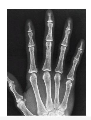
- Pathognomonic Features
- At 2nd& 3rd proximal / mid phalanges on Radial aspect: Subperiosteal resorption
- In skull :Salt & pepper skull appearance
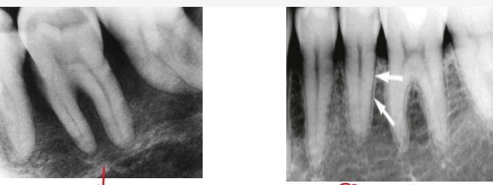
Loss of lamina dura Normal lamina dura
- Multifocal irregular lytic lesions scattered throughout body in metaphysis k/a “Brown tumors/Osteitis cystica fibrosa”
Important Information
- Brown tumors : Bleeding occurs within them resulting in hemosiderin deposition
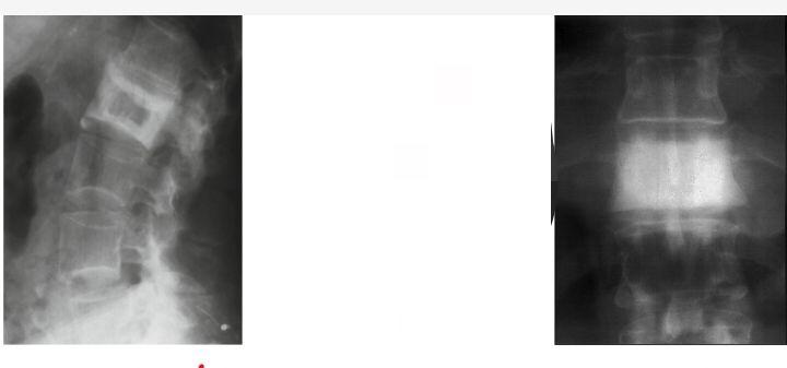
Secondary hyperparathyroidism
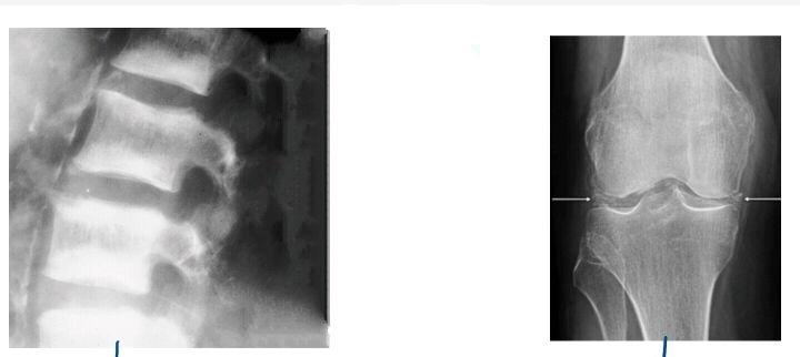
- Chronic kidney disease →↓Ca2+, ↑PO3 → HPT (2°)
- Hallmark finding : Sclerosis
- Alternative bands of sclerosis & osteopenia: Rugger jersey spine
- Knee & wrist : Calcification of meniscus k/a chondrocalcinosis
Important Information
- Chondrocalcinosis
- 2°HPT
- Pseudo gout (due to calcium pyrophosphate deposition)
- Wrist : Triangular fibrocartilage calcification
- Knee: Meniscus calcification
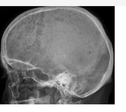
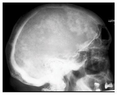
Paget’s Disease
- May have visual loss, hearing loss (because cranial nerves getting compressed)
- Sclerotic phase
- Skull markedly enlarged with multiple sclerotic spots :Cotton wool skull
- ↑Hat size: Tam O’ Shanter sign
- Lytic phase
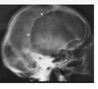
- Osteoporosis circumscripta [large area of decreased bone density]
- In long bones
- Lysis with advancing edge :Blade of grass / candle flame sign
- Spinal manifestation
- Peripheral sclerosis, central osteopenia: Picture frame vertebra
- Ivory vertebra
Important Information
- Ivory vertebra
- Lymphoma
- Hodgkin
- Sclerotic metastasis
- Prostate : male
- Breast : female
- Paget’s disease
- In case of treated TB
- Lymphoma
- In bone scan
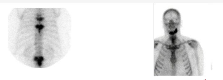
- Tc99m with MDP (Methyl Diphosphonate) [Picks up new bone formation area]
- “Mickey Mouse sign” in vertebra
- “Lincoln sign” in mandible
Mickey Mouse Sign
- USG LL → Sapheno femoral junction [GSV, SFV, SFA]
- USG Antenatal→ Anencephaly
- Bone scan → Paget’s disease
- Brain MRI → Midbrain atrophy in case of PSNP
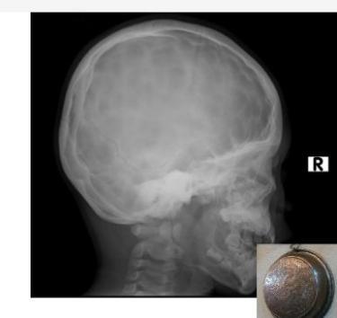
Acromegaly
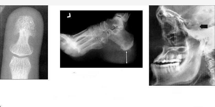
- Due to increased growth hormone (after growth plate fusion)
- Over growth in distal phalanges : “Spade phalanx”
- Gross increase in heel pad thickness
- > 23 mm in female
- > 25 mm in male
- In skull
- Widened sella
- Prognathism
- Enlarged sinuses
- Thick bones, PNS
.jpg)
Langerhans Cell Histiocytosis

- Multisystemic disorder
- Child with multiple bone swelling, multiple lymph nodes, multiple skin lesions
- In skull
- Lytic lesion: Geographical with beveled margin (differential destruction of outer & inner table)
- Spine
- One of the Vertebra completely destroyed :“Vertebra plana”
Important Information
- LCH : MC cause of vertebra plana in children
- Ewing sarcoma, lymphoma, leukemia can also cause vertebra plana
- Mandible
- “Floating teeth sign” (due to multiple erosion in mandible)
Radiology Related Articles:
Multiple Myeloma
- Multiple punched out lesion Aka Raindrop appearance [Also seen in lytic metastasis]
Sturge – Weber syndrome
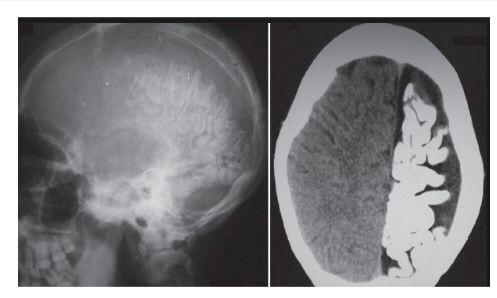
- Complete cerebral atrophy with tram track calcification which leads to refractory seizure
- Port wine stain
- MC affected: Ophthalmic branch of Trigeminal nerve
- Congenital glaucoma (+)
Hemolytic Anemia
- Sickle cell anemia, Thalassemia
- Hair on end / Crew cut appearance
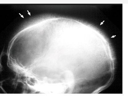
Raised ICP
- Copper beaten sign
- Earliest sign on X-ray
- Children: Sutural diastasis (Sutural widening )
- Adults : Dorsum sella erosion
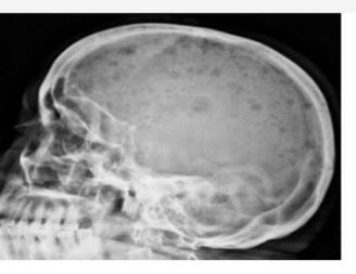
Osteopetrosis
- Congenital dysplasia
- Child with hepatosplenomegaly having pancytopenia
- Diffuse ↑ density
- Bone within Bone appearance / Sandwich sign
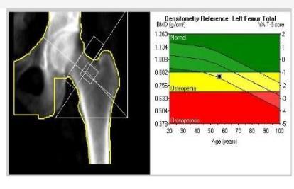
Osteoporosis
- Diffuse ↓ density
- End plates compressed :“Codfish appearance ” [due to weak bone & weight of the body]
- Important Information
- Codfish appearance also seen in Osteomalacia (Vit D def. in adults)
- IOC: Dexa Scan (Dual energy X-ray absorptiometry)
- If we compare the bone mineral density of the patient with young adult : T score
- If we compare the bone mineral density of the patient with the same aged person : Z-score
- WHO scoring used T-score
- +2 to -1 : Normal
- -1 to -2.5 : Osteopenia
- < -2.5 SD : Osteoporosis
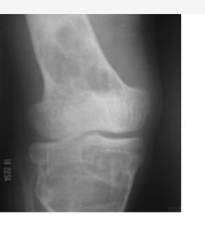
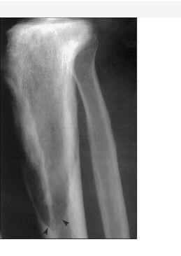
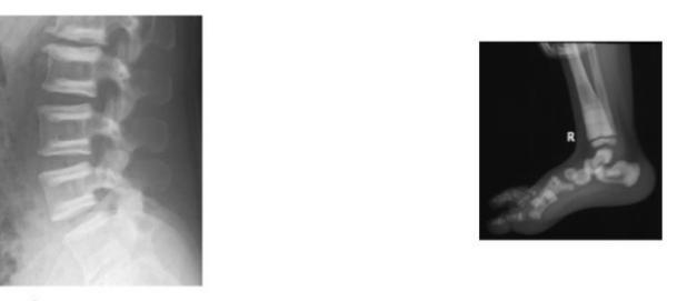
Rickets
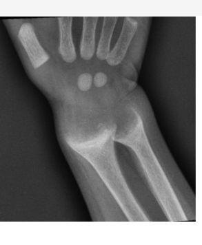
- Child with widening of joints predominantly wrist, ankle
- Vit D deficiency
- Earliest finding: Increased growth plate / Zone of provisional calcification
- Due to decreased vitamin D →↓Ca2+
- Unmineralized osteoid accumulates near growth plate
- Seen in Metaphysis: Cupping, Splaying, Fraying (irregularities)
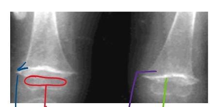
- Sign of healing rickets

- White metaphyseal line
Important Information
- White metaphyseal line
- Healing rickets
- Lead poisoning
- Treated Leukemia
- Scurvy
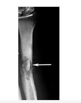
Scurvy

- Vitamin C deficiency
- Bone density decreased
- Frenkel’s white line
- Wimberger sign [Epiphyseal sharp margin]
- Pelkan spur (metaphyseal projection)
- Trummerfeld zone (Translucent zone above white line)
- Subperiosteal hemorrhage
- Very painful [Scurvy is known as Pseudo paralysis state]
Chronic Osteomyelitis
- Central white dead bone (no demineralization) → Sclerotic k/a Sequestrum
- Surrounding translucent granulation → Involucrum
- Defect through which the pus comes out → Cloaca (then will have draining sinus)
To study this topic in detail along with other high-yield radiology topics, download the PrepLadder app and discover engaging video lectures by expert faculty.

PrepLadder Medical
Get access to all the essential resources required to ace your medical exam Preparation. Stay updated with the latest news and developments in the medical exam, improve your Medical Exam preparation, and turn your dreams into a reality!
Navigate Quickly
Secondary hyperparathyroidism
Paget’s Disease
Mickey Mouse Sign
Acromegaly
Langerhans Cell Histiocytosis
Multiple Myeloma
Sturge – Weber syndrome
Hemolytic Anemia
Raised ICP
Osteopetrosis
Osteoporosis
Rickets
Scurvy
Chronic Osteomyelitis
Top searching words
The most popular search terms used by aspirants
- NEET PG Radiology
- NEET PG Strategy
PrepLadder Version X for NEET PG
Avail 24-Hr Free Trial