Radiotherapy Types | Cancer Treatment with Radiation
Mar 30, 2023

- Therapy is given using Radiation.
- Radiation is a ‘Double-edged sword’
- It kills cancerous cells; it may also damage normal cells.
- Targets cancerous cells and prevents damage to normal cells.
- It kills cells by damaging DNA which produces free radicals.
In this blog, you’ll get a brief overview of this important radiology topic for NEET PG exam preparation. Read on.
Radiotherapy Types
- It depends on the source of radiation.
- Two types:
- Teletherapy: EBRT (External Beam Radiotherapy), source is at a distance,
- Brachytherapy: source is nearer to the patient.
- Brachytherapy is divided into 3 types:
- Interstitial (into the tissue): Ca Prostate, Ca Breast
- Intracavitatory (accessible cavity): Accessible tumors like Ca Cervix
- Mold (Superficial cancers): Penis, Eye cancers
| Golden Points Disadvantage of Teletherapy as compared to Brachytherapy is the beam coming from outside, the target part along with adjacent parts are also exposed Brachytherapy reduces the risk of exposure of adjacent parts to radiation. In Teletherapy, Cobalt machine and LINAC (Linear Accelerator) are used. |
- The Cobalt machine produces an artificial radioisotope (Co60).
- Half-life of Co60: 5.2 years
- It decays into Ni60.
- LINAC does not need any radioisotope, the fast moving electron beam generates radiation used for radiotherapy.
- X-rays generated by LINAC have various energies. They are:
- Ortho voltage < Super voltage < Mega voltage
- Penetration is directly proportional to energy.
- Mega voltage has higher energy and penetration power.
- Ortho voltage has lower energy, can be used in superficial cancers and IORT (Intraoperative Radiotherapy).
- Eg: A patient with carcinoma of pancreas, tumor is removed and the residual cells are to be killed which are superficial. Less energy or less permeable rays are enough. Hence, electron beam or ortho voltage is used.
| Remember Whole body EBRT is used in Mycosis fungoides. |
LINAC Machine
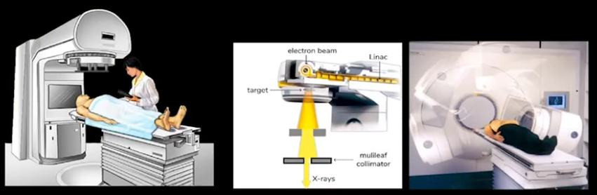
- Overhead x-ray tube which emits x-ray beams.
- It also has a robotic arm, which moves around the patient.
- No frame is used to immobilize the patient.
- Frameless radiotherapy.
Interstitial Brachytherapy
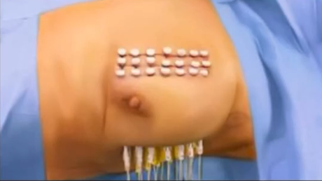
- Interstitial Brachytherapy has no cavity.
- To insert the radioactive source, After loading is the technique to be followed.
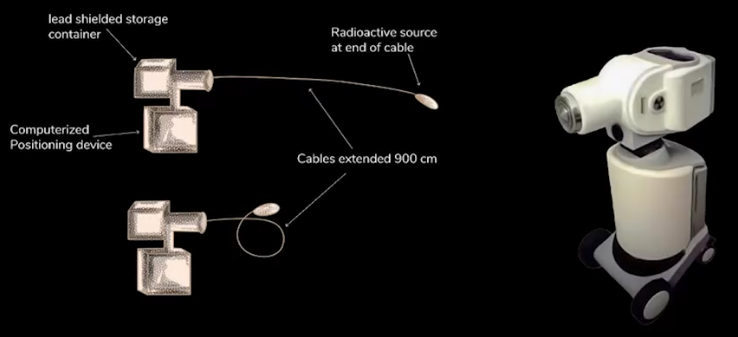
Afterloading
- Empty shell is put into the tissue, through which a robotic arm, a Lead-shielded storage container, sends the cable and automatically loads the shell.
- Reduces the manual handling of radioactive materials.
Agents used for radiotherapy
- X-rays (LINAC) are most commonly used.
- Proton beams, neutrons, Gamma and beta rays are also used.
- These pure emitters are used in Systemic Brachytherapy, where the isotope diffuses and reaches the site of action.
- Seed Brachytherapy involves the localized action of the isotope.
- Phosphorus 32 (P32) is used in treatment of polycythemia vera.
- Strontium is used for metastatic bone pain.
- Iodine 131 (I131) is a Beta emitter (not pure form). It emits gamma radiations as well.
- Gamma radiation can be used in Systemic Brachytherapy.
- Beta rays stay in the body and show the therapeutic effects, gamma rays exit the body.
- I131: Half-life is 8 days.
- Beta, Gamma rays are emitted from the nucleus of the atom.
- X-rays are extranuclear.
.jpg)
Bragg peak
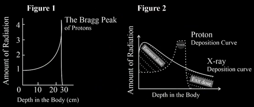
- In a proton beam, velocity is inversely proportional to dose given to the body.
- As the beam travels deeper into the body, the velocity decreases and the dose increases suddenly.
- Superficial structures are protected when proton beams are used.
- Targeted pencil beams like radiotherapy can be given.
- It affects only the tumor, surrounding cells are protected.
- Example: A child with a brain tumor is given proton beam therapy, because the surrounding cells are not to be affected.
Image of comparison between X-ray with Bragg peak
- X-rays give the maximum dose on the body.
- Most common side effect of radiotherapy is skin erythema.
Comparison between X-ray with bragg peak in medulloblastoma

- In the proton beam, localized action is given.
- In X-rays the normal cells are also affected.
The half-life of Radioisotopes
Isotope Uses Half-life Iodine-123 Diagnosis 13 hours Iodine-125 Permanent implant in Ca Prostate 60 days Iodine-131 Systemic Brachytherapy (Grave’s disease, Thyroid cancer) 8 days Technetium-99m Diagnostic radioisotope in Scintigraphy 6 hours Fluorine-18 Diagnostic radioisotope in PET 2 hours (110 mins) Radium-226 1600 years Cesium-137 30 years Cobalt-60 5.26 years Iridium-192 74.2 days Iodine-125 Permanent implant in Ca Prostate 60.2 days Golden Points
I-127 is a naturally occurring radioisotope and has no radioactivity.
Radium-226, has the highest half-life.
First radioisotope used in humans was Radium-226.
It produces Radon (decay compound), which causes carcinoma of lungs.
It emits alpha, beta, gamma rays.
- Radioisotopes with shorter half-lives can be used as permanent implants in Brachytherapy.
- These decay on their own.
- Radioisotopes with longer half-lives can be used as temporary implants in Brachytherapy.
- These implants are to be removed.
Also Read: NEET PG: High-Yield Topics For Radiology
Techniques Used to Reduce the Damage to Normal Tissues
- Gating
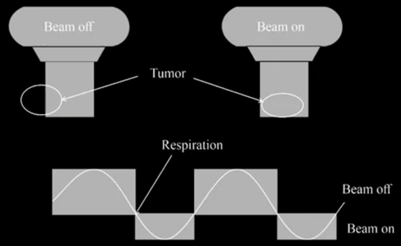
- Respiratory gating is used in lung cancer to protect normal tissue.
- The tumor moves with respiration.
- Radiation is produced only when the tumor is in the line of the beam.
- If a tumor goes off the beam line, radiation stops.
Image of 3DCRT, IMRT, VMAT, IGRT 21.55 mins
- 3DCRT
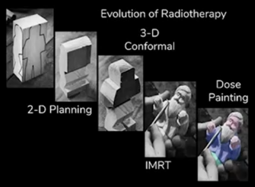
- 3-Dimensional Conformal Radiation Therapy
- The radiation is exposed according to the shape of the tumor.
c. IMRT
- Intensity Modulated Radiation Therapy
- It is better than 3DCRT.
- Used for tumors with irregular shape
- Multiple small beams of various intensities are exposed on the tumor cells.
- This cannot be used in lung cancer.
d. VMAT
- Volume Modulated Arc Therapy
- Radiation beam rotates around the tumor and changes intensity.
e. IGRT
- Image Guided Radiation Therapy
- Radiation is exposed under image guidance.
f. Gamma knife
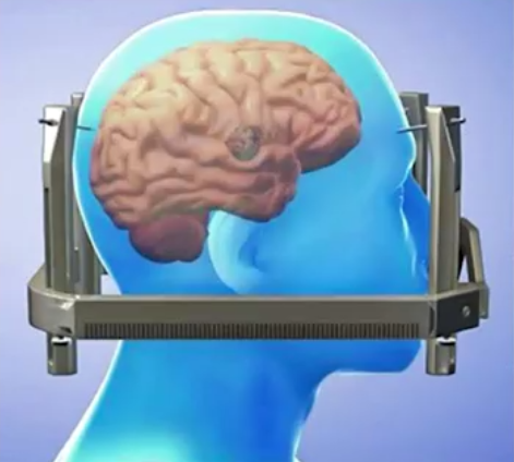
- It comes under Stereotactic radiotherapy or surgery (SRS).
- It was invented by neurosurgeon Lars Leksell.
- Gamma rays are used to remove the tumor cells.
- Used in brain lesions like
- Vestibular schwannoma
- Trigeminal neuralgia
- AV malformations
- Pituitary adenoma
- Meningioma
- Not used in TB.
- Leksell's frame prevents immobilization of the patient's head.
- MRI is done to localize the brain lesion.
- Gamma knife emits multiple radiations over the coordinates placed on the tumor site.
- Rays get focussed on the tumor site and ablated.
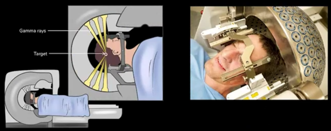
Gamma knife
- Disadvantage of the Gamma knife is that the frame is required.
g. Cyberknife
- Stereotactic Body Radiotherapy (SBRT)
- It uses x-rays (LINAC)
- Frameless
- Used in whole body
- Used for Localized early stage cancers.
- Not used in
- Ca tongue, as it has lymph nodes (metastasized).
- Ca Breast, with lymphangitis carcinomatosis.
h. NCT
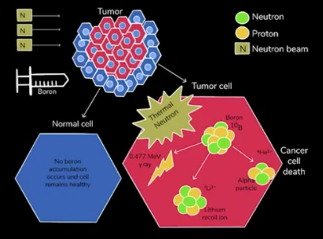
- Neutron Capture Therapy
- Used for brain tumors
- Boron is injected (BPA), which is taken up by tumor cells and not healthy cells.
- Thermal neutron is exposed, which is captured by Boron.
- This interaction generates alpha particles, which kills the cell.
Also Read: Musculoskeletal Radiology: Bone Tumors
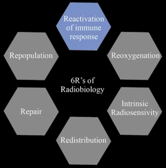
6 R's of Radiobiology
i. Fractionated Radiotherapy
- A single high dose may be given as small fractions of low doses.
- Single high dose may damage normal cells also.
- Advantages: 6 R's of Radiobiology
- Reoxygenation
- Repopulation
- Repair
- Redistribution
- Intrinsic Radiosensitivity
- Reactivation of immune response
1. Repair
- Fractionated doses do not damage normal cells
- If damaged they get time to repair sublethal damage of normal cells.
- Fractions of doses are given 5 days per week and weekends are off.
2. Reoxygenation
- Hypoxic cells are not killed by radiotherapy, due to the free radicals.
- New capillaries are formed during the gap, which provides oxygen.
| Golden Point: Oxygen acts as a Radiosensitizer. |
- Radiation Sensitizers
- Most potent: Hyperbaric oxygen
- Metronidazole
- Chemotherapeutic drugs
- 5-FU
- Cisplatin
- Radio Protectors
- Amifostine protects all parts of the body except the brain (does not cross BBB).
- Sulfhydryl containing groupys (Oxygen scavengers)
- Cysteine
- Pentoxiphylline
- Chlorhexidine-stomatitis
- Radiation Potentiators
- Anthracyclines
| To Remember: Anthracyclines may show radiation recall syndrome |
Golden Points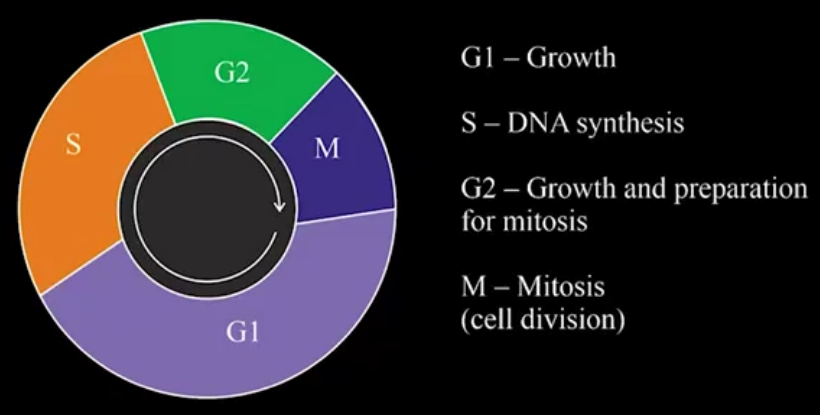 According to the Law of Bergonie, the more the cells that are rapidly dividing, the more they are sensitive to radiation. According to the Law of Bergonie, the more the cells that are rapidly dividing, the more they are sensitive to radiation. M-phase (dividing phase) is most radiosensitive phase S-phase (Synthesizing phase) is most radioresistant phase |
3. Redistribution
- It is based on the cell cycle phase of cancer cells.
- Cells on the 1st day are in S phase, and no action of radiation is seen.
- On the next day they are in M phase.
- In the M phase, the cells are rapidly killed.
4. Repopulation
- Double-edged sword.
- All the cancerous cells are not killed. They may repopulate the next day.
5. Intrinsic Radiosensitivity
|
Golden points
|
6. Reactivation of the immune response
- Activates the immune response
Also Read: Why is Radiology the most preferred branch?
Types of Fractionated Radiotherapy
Standard
- Mon-Fri daily one dose
- Weekend off
Hyperfractionated
- CHART (Continuous Hyper Fractionated Accelerated Radiotherapy) regime.
- Treat Carcinoma of lungs, Non-small cell cancers like adenocarcinoma.
Hypofractionated
- Given in Palliative treatment
Craniospinal Irradiation
- It is given prophylactically in tumors which have drop metastasis.
- Medulloblastoma, drops to the spine and causes spinal tumors like sugar coat (Zuckerguss in meninges with metastasis).
- ALL (Acute Lymphoblastic Leukemia)
- Small cell carcinoma of lungs
Previous Year Questions
Q. What does LINAC produce?
Ans: LINAC produces X-rays and electrons.
Q. What is the most common radiation used in IORT?
Ans: Electron beam or ortho voltage, as it has less penetration power.
Q. Afterloading is used in?
Ans: Brachytherapy
Q. Which radioisotopes are pure beta emitters?
Ans: Phosphorus 32 (P32), Strontium, Yttrium.
Q. Which is shown in Bragg peak?
Ans: Proton beam
Q. Which Radioisotopes are used as permanent implants in Brachytherapy?
Ans: C I G Y R P
- Cesium-131
- Iodine-125
- Gold
- Yttrium
- Radon-222
- Palladium
Q. A child undergoing radiotherapy, which tissue may be least affected?
Ans: Nervous tissue
Q. What is the first hormone deficiency in Craniospinal Irradiation?
Ans: Growth hormone
Download the PrepLadder app now and unlock a 24-hour FREE trial of premium high-yield content. Access Smarter Video Lectures, Game Changing Qbank, Audio QBank, Structured Notes, Treasures, and Mock Tests for FREE to ace your NEET PG preparation. Elevate your study experience and gear up for success. Start your journey with PrepLadder today!

PrepLadder Medical
Get access to all the essential resources required to ace your medical exam Preparation. Stay updated with the latest news and developments in the medical exam, improve your Medical Exam preparation, and turn your dreams into a reality!
Navigate Quickly
Radiotherapy Types
LINAC Machine
Interstitial Brachytherapy
Afterloading
Agents used for radiotherapy
Bragg peak
Image of comparison between X-ray with Bragg peak
Comparison between X-ray with bragg peak in medulloblastoma
The half-life of Radioisotopes
Techniques Used to Reduce the Damage to Normal Tissues
c. IMRT
d. VMAT
e. IGRT
f. Gamma knife
g. Cyberknife
h. NCT
6 R's of Radiobiology
i. Fractionated Radiotherapy
1. Repair
2. Reoxygenation
3. Redistribution
4. Repopulation
5. Intrinsic Radiosensitivity
6. Reactivation of the immune response
Types of Fractionated Radiotherapy
Standard
Hyperfractionated
Hypofractionated
Craniospinal Irradiation
Previous Year Questions
Top searching words
The most popular search terms used by aspirants
- Medical PG Preparation
- NEET PG study tips
PrepLadder Version X for NEET PG
Avail 24-Hr Free Trial