Breast Imaging Reporting And Data System - NEET PG Surgery
Mar 28, 2023

Carcinoma of the breast is a type of cancer that arises from the cells in the breast tissue. It is one of the most common types of cancer that affects women, and the incidence is increasing globally. Breast Imaging Reporting and Data System (BI-RADS), a standardized system is used by radiologists to classify breast imaging findings and provide recommendations for follow-up.
For the NEET PG exam, it is essential to understand the various aspects of carcinoma breast, including its diagnosis, management, and treatment options. Furthermore, understanding the contraindications for BCS in carcinoma breast is crucial for making informed decisions about patient management and treatment.
Read this blog further to get a quick overview of this important surgery topic for NEET PG exam preparations.

Breast
Causes and Types of Nipple Discharge
|
ETIOLOGY |
||
|
Bloody Nipple Discharge |
Serous Nipple Discharge |
Greenish/Blackish/Grumous/Pultaceous Nipple Discharge |
|
|
|
Treatment
- Duct papilloma is a benign condition: Microdochectomy (Excision of involved duct only)
- Duct Ectasia: Hadfield's Operation (Conical Excision of involved duct)
Carcinoma Breast
- Generally, adenocarcinomas are more common in high socio-economic status and squamous cell carcinomas are more common in low socio-economic status
- Carcinoma Breast is adenocarcinoma, Most common in females belonging to high socioeconomic status
Risk Factors of Carcinoma Breast
- Advancing age
- Western countries
- High socio-economic status
- Alcohol intake
- High fat diet leading to obesity
- State of hyper-estrogenemia are
- Early menarche
- Late Menopause
- Nulliparity
- Late first full-term pregnancy
- Positive family history specially from maternal side
- Genetic mutation
- BRCA-1: Mainly for female Breast-Carcinoma (Ladies First)
- BRCA-2: Mainly for male Breast-Carcinoma
- Personal history of malignancy
- Ovarian Carcinoma
- Endometrial Carcinoma
- Hormone replacement therapy
- Therapeutic radiation exposure
Important Information
- OCP and Smoking are not the significant risk factors for Ca Breast
- Long duration of breast feeding is protective for Ca Breast.
Ductal Carcinoma Insitu(DCIS)
- Though ducts are present in males and females it is common in both
- It can progress to invasive ductal cancer
- On the basis of nuclear grade and necrosis ductal carcinoma insitu divided into
- Low grade
- High grade
| Low grade | High grade |
| Cribriform | Solid |
| Papillary | Comedocarcinoma |
| Micropapillary | - |
Investigations
- Sensitive investigation for diagnosis of microcalcification in DCIS is Mammography
- Sensitive investigation for diagnosis of DCIS is MRI
Treatment
- Non-palpable DCIS: Excision by needle localization with specimen mammography
- Low grade DCIS: Lumpectomy
- DCIS with limited disease: Lumpectomy + Radiotherapy
Lobular Carcinoma INSITU
- Lobules are present only in females so lobular carcinoma insitu is present only in females
- It arises from terminal duct lobular units
- It is multicentric and bilateral therefore there is increased risk of bilateral breast cancer
Pathology
- Cytoplasmic mucoid globules
- Hallmark: Indian file pattern
Clinical features
- Lump with ill-defined margins
Diagnosis
- Neighborhood calcification
Treatment
- Observation
- Chemoprevention: Tamoxifen, Raloxifene
- Prophylactic B/L mastectomy
- Important Information
- Most common Histological type: Adenocarcinoma
- Most common Histological subtype: Invasive ductal cancer
- Least common Histological subtype: Papillary
- Most malignant Breast Carcinoma: Inflammatory Breast Carcinoma - (worst prognosis)
- Least malignant Breast Carcinoma: Tubular (best prognosis)
- Most common Site of breast cancer: Upper Outer Quadrant (Max Amount of Breast tissue)
- Least common area of Breast Carcinoma: Lower Inner Quadrant
- Breast Carcinoma is more common in Left Breast
- Most common route of spread in breast cancer is Lymphatics
- Most common site of Metastasis is BONES (Lumbar Vertebra > Femur > Thoracic vertebrae- "LFT")
- Carcinoma Breast is most common Malignancy causing Osteolytic & Osteoblastic secondaries in bones (Osteolytic >Osteoblastic)
- Most common cause of Death in Carcinoma Breast is Malignant pleural Effusion
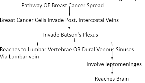
- Most common primary for Leptomeningeal Metastasis is Carcinoma breast
- Most common primary for Brain Metastasis is Carcinoma Lung > Carcinoma Breast
Clinical features
- Most common presentation is Lump
- In advanced cases
- Architectural distortion
- Asymmetry
- Skin fixity
- Fixity to Chest Wall
- Involvement of nipple: Nipple Retraction / Deviation / Ulceration

Signs and Symptoms of Metastasis
- Backache
- Headache
- Dyspnea
- Jaundice
- Anorexia
- Weight loss
- PEAU-D-ORANGE: Most Conspicuous Sign of Breast Ca, and it is due to cutaneous edema caused by permeation of lymphatics by tumor cells.
- Skin depression/ Dimpling
- Puckering (Fold/ wrinkling of Skin)
- Both of these are due to involvement of ligament of cooper
- Cancer - En - Cuirasse: Presence of multiple nodules and ulceration in the breast due to involvement of breast skin and chest wall.
Triple Assessment in Ca-Breast
- It has positive predictive value (PPV): 99.9%
- Components of Triple Assessment are
- Clinical: Signs & Symptoms
- Imaging: Mammography/ USG
- Tissue Sampling: Biopsy/ FNAC
Also Read:
- Abnormalities in SMALL INTESTINE - NEET PG Surgery
- Bartholin Cyst: Causes, Symptoms, Diagnosis, Treatment, Prevention and Complications
- Hypospadias: Causes, Symptoms, Risk Factors, Diagnosis, Treatment and Complications
- Bakers Cyst: Causes, Symptoms, Diagnosis, Treatment, Prevention and Complications
Investigations
- 1st Investigation done in suspected case of Breast Ca: Mammography
- Investigation of choice for diagnosis of Carcinoma breast: Biopsy (true cut biopsy / core cut biopsy/ Needle biopsy)
Mammography
- Investigation of choice for Screening of Carcinoma Breast
- Age: 45 years (After that Annual Mammography)
- Bremsstrahlung X-rays are used
- Radiation Exposure: 0.1 Centi Gray/Study
- 1 Mammography = 4 Chest X-rays
Reading of Mammography
- Look for nipple: Normal or Retarded
- Look for skin
- Focus on opacity-Calcification: Microcalcification (malignancy) & macrocalcification (benign)
- The space between pectoralis major and breast tissue is retro mammary space
Differentiation of Benign and Malignant Lesion on Mammography
|
Benign |
Malignant |
|
|
Opacity |
|
|
Calcification |
|
|
Associated changes |
Present

|


BIRADS (Breast Imaging Reporting And Data System)
|
Category |
Description |
|
0 |
Incomplete Assessment, Additional Imaging is required: risk of malignancy NA |
|
1 |
Negative, Annual Mammography recommended: 0% risk of malignancy |
|
2 |
Benign, Annual Mammography recommended: 0% risk of malignancy |
|
3 |
Probably Benign, Short term follow up:> 0-2% risk of malignancy |
|
4 |
Suspicious of Malignancy
|
|
5 |
Highly suggestive of Malignancy, Intervention is recommended:> 95% risk of malignancy |
|
6 |
Biopsy Proven Malignancy |
Role of MRI in Breast Cancer
- Investigation of choice for Screening of Breast Carcinoma in High-risk females with family history and BRCA mutation
- Investigation of choice for Implant Related Complications
- Investigation of choice to differentiate scar from benign lesions
USG
- 1st investigation in female < 35 years with lump
Important Information
- Young female < 35 years have dense and glandular breast → which decrease the sensitivity of Mammography
PET Scan
- Investigation of choice for diagnosis of distant metastasis
- Investigation of choice to differentiate scarring, necrosis, fibrosis from recurrence
Bone Scan
- Investigation of choice for diagnosis of Bone metastasis
BCS (Breast Conservative Surgery)
- Done for early invasive breast cancer: I, IIA, IIB
- Lumpectomy: Excision of lump
- Wide local excision with 1 cm margin
- Quadrantectomy: Excision of whole quadrant
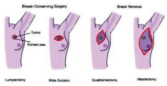
- In all patients of Breast Conservative Surgery we give Radiotherapy (if there is contraindication to Radiotherapy then there is contraindication to Breast Conservative Surgery)
Contraindication for Breast Conservative Surgery
Absolute Relative Pregnancy H/o Collagen Vascular DiseaseSclerodermaActive lupus erythematosus 2 or > 2 tumors in different quadrants or diffuse malignant appearing microcalcifications Multiple tumors in the same quadrant or indeterminate calcification Persistently positive margins Large tumor in small Breast H/o Therapeutic radiation exposure Large pendulous breast Centrally located tumor
In early invasive breast cancer if there is contraindication for Breast conservative Surgery, we do simple or total mastectomy
To study this topic in detail along with other important topics from Surgery, download the PrepLadder app and get access to in-depth video lectures, study notes and MCQs for practice.

PrepLadder Medical
Get access to all the essential resources required to ace your medical exam Preparation. Stay updated with the latest news and developments in the medical exam, improve your Medical Exam preparation, and turn your dreams into a reality!
Navigate Quickly
Breast
Causes and Types of Nipple Discharge
Treatment
Carcinoma Breast
Risk Factors of Carcinoma Breast
Ductal Carcinoma Insitu(DCIS)
Investigations
Treatment
Lobular Carcinoma INSITU
Pathology
Clinical features
Diagnosis
Treatment
Clinical features
Signs and Symptoms of Metastasis
Triple Assessment in Ca-Breast
Investigations
Mammography
Reading of Mammography
Differentiation of Benign and Malignant Lesion on Mammography
BIRADS (Breast Imaging Reporting And Data System)
Role of MRI in Breast Cancer
USG
PET Scan
Bone Scan
BCS (Breast Conservative Surgery)