Last 5-Year PYQs in Radiology for FMGE
Aug 12, 2025

When it comes to preparing for FMGE, there is no smarter way to understand exam patterns, identify high-yield topics, and refine your strategy than solving previous years’ questions (PYQs).
Radiology, being a high-yield and image-intensive subject, plays a crucial role in your FMGE success. It covers X-ray, CT, MRI, ultrasound, nuclear medicine, and interventional radiology — making both visual recognition and conceptual clarity essential.
But what if you had access to the most frequently asked questions from the last five years? That’s exactly what this blog offers.
We’ve compiled high-yield Radiology PYQs that have been repeatedly tested in FMGE, along with detailed explanations to help you revise efficiently, correct common errors, and sharpen your image-based question-solving skills.
Download FMGE Last 5-Year PYQs – Subject-wise PDFs
|
|
|
|
|
|
|
|
|
|
|
|
|
|
|
|
|
|
|
|
|
|
|
|
|
|
||
Let’s dive in and supercharge your Radiology prep for FMGE 2025!
Q1. A patient presents with a neck mass and laboratory findings of increased T4 and decreased TSH. The patient has a history of I-131 ablation therapy. Which of the following is a likely side effect of the treatment?
- Acute Thyroiditis
- Hypothyroidism
- Hyperthyroidism
- Thyroid Storm
Answer: 3) Hyperthyroidism
Q2. Which of the following methods is used to measure fetal anemia?
- Umbilical artery Doppler
- Cord blood sampling
- Chorionic villus sampling
- Peak systolic velocity in the fetal middle cerebral artery (MCA)
Answer 4: Peak systolic velocity in the fetal middle cerebral artery (MCA)
Q3. A 70-year-old patient presents with absolute constipation and abdominal distension. And the X-ray abdomen is given below. What is the most likely diagnosis?
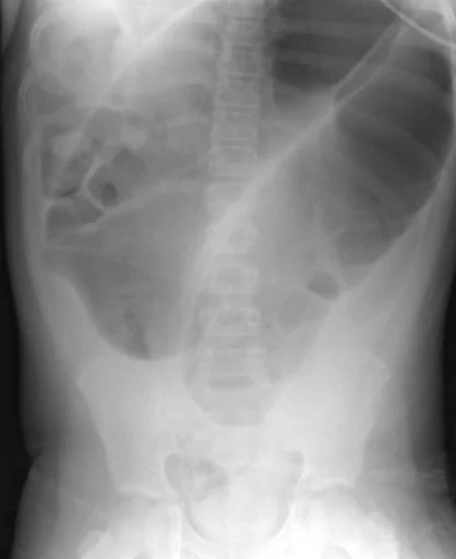
- Caecal Volvulus
- Sigmoid Volvulus
- Intestinal Obstruction
- Small Bowel Volvulus
Answer 2: Sigmoid Volvulus
Q4. Which of the following is a primary use of this imaging modality?
- Staging of esophageal cancer
- Evaluation of gastroesophageal reflux disease (GERD)
- Identifying the cause of dysphagia
- Assessing cardiac and aortic pathology
Answer 1) Staging of esophageal cancer
Q5. What is the investigation shown in the image?
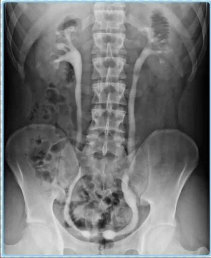
- Intravenous Pyelogram (IVP)
- Retrograde Pyelogram
- Computed Tomography (CT) Urography
- Magnetic Resonance Urography
Answer 1: Intravenous Pyelogram (IVP)
Q6. A 55-year-old female patient presents to the emergency department with chills and rigors. She has acute pain in right flank region. The imaging is shown below. What is the most likely diagnosis?
- Cholecystitis
- Emphysematous pyelonephritis
- Small bowel obstruction
- Volvulus
Answer: B - Emphysematous pyelonephritis
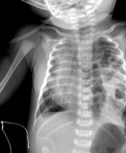
Q7. A newborn presents with bilious vomiting. Which sign is depicted in the following radiograph?
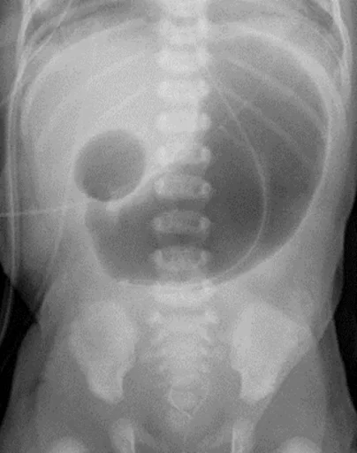
- Double bubble sign
- Air crescent sign
- Cupola sign
- Triple bubble sign
Answer A - Double bubble sign
Q8. A patient presented with a head injury after a hammer fell on the skull. CT is shown below. What is the likely diagnosis?
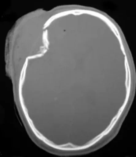
- Concussion
- Epidural hematoma
- Depressed skull fracture
- Subarachnoid hemorrhage
Answer: C - Depressed skull fracture
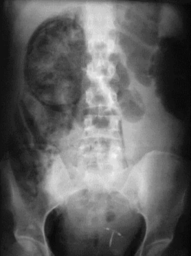
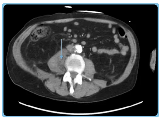
Q9. A 28-year-old patient presents with recurrent urinary tract infections and left flank pain. CT scan of the abdomen was performed and the image is shown below. What is the likely diagnosis?
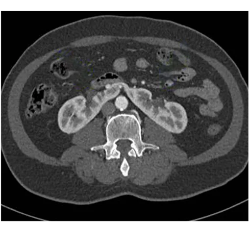
- Horseshoe kidney
- Polycystic kidney disease
- Renal ectopia
- Hydronephrosis
Answer: 1 - Horseshoe kidney
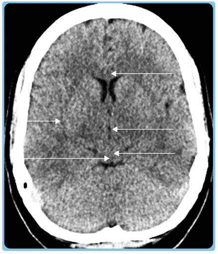
Q10. Identify the pathology in the shown CT:
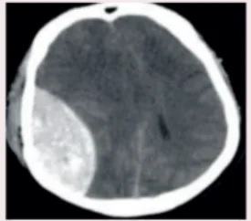
- Intraventricular bleed
- Massive Epidural hemorrhage
- Subdural hemorrhage
- Subarachnoid hemorrhage
Answer B: Massive Epidural hemorrhage
Q11. Identify the sign?
- Claw sign
- Target sign
- Coffee bean sign
- Lead pipe sign
Answer B: Target sign
Q12. Which of the subsequent investigations operate based on the same principle?
- CT and MRI
- CT and X-ray
- USG and HIDA Scan
- MRI and PET Scan
Answer: B CT and X-ray
Q13. Investigation of choice for GERD:
- USG
- HIDA
- Manometry
- 24 hour pH monitoring
Answer: D 24 hour pH monitoring
Q14. What is the probable diagnosis for a 36-year-old farmer who presented with a cough, chest pain lasting one week, fatigue persisting for a month, and a chest x-ray revealing the image provided below?
- Hydatid cyst
- Anthrax
- Silicosis
- Byssinosis
Answer: A) Hydatid cyst
Q15. A 13-year-old boy presents to you with a limitation of abduction and internal rotation of the left hip. The x-ray appearance of his left hip joint is shown in the below image. What is the diagnosis of this patient?
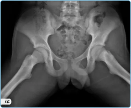
- Slipped capital femoral epiphysis
- Perthes disease
- Development of dysplasia of the hip
- Ankylosing spondylitis
Answer: A) Slipped capital femoral epiphysis
Q16. What is the likely diagnosis for a 20-year-old male who presents with pain in the right flank, along with fever and chills? The patient experiences pain when the hip is passively extended on the right side. Lab tests indicate an increased white blood cell count, and a CT scan reveals specific findings.
- Pyelonephritis
- Psoas abscess
- Appendicitis
- Torsion of right undescended testis
Answer: B) Psoas abscess
Q17. Identify the labelled structure in the image?
- Midbrain
- Pons
- Medulla
- Cerebellum
Answer: A) Midbrain
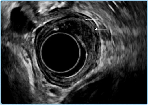
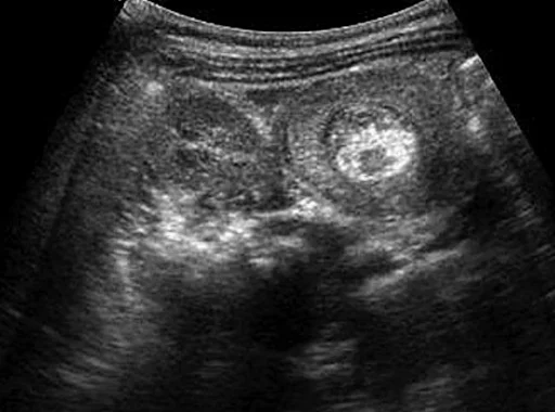
Q18. Please identify the structure labelled 'X' in the given ultrasound image of the liver.
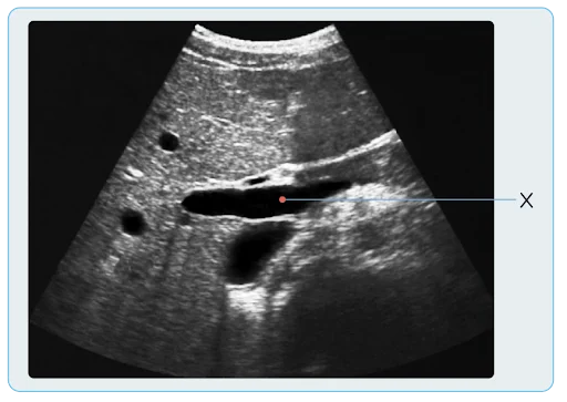
- Portal vein
- Hepatic artery
- Hepatic vein
- Inferior vena cava
Answer : A) Portal Vein
Q19. Please provide your analysis of the diagnosis based on the CT scan of the chest displayed below.
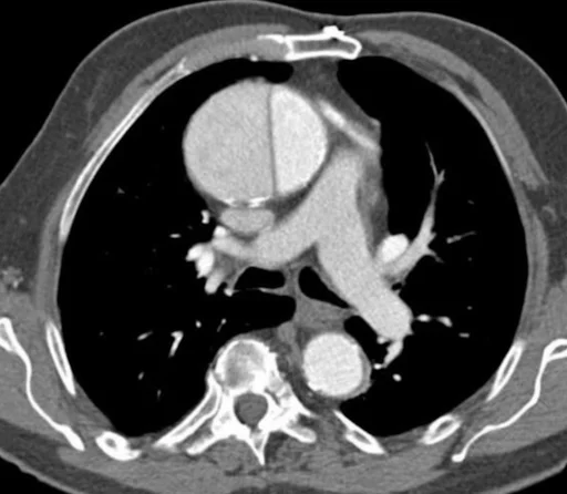
- Aortic dissection
- Pulmonary embolism
- Cardiac myxoma
- Aortic aneurysm
Answer: A - Aortic dissection
Q20. A 55 year old male patient presented with complaints of dyspnea with congestive heart failure . The clinician wants to know whether it's HF with preserved EF or reduced EF. Which of the below investigations is used for calculation of ejection fraction ?
- MUGA
- SPECT using thallium 201
- PET myocardial perfusion imaging
- Sestamibi scan with pharmacological stress
Answer A: MUGA
Q21. Which of the following images accurately represents the medical condition described in the given scenario?
- Diaphragmatic hernia
- Intestinal obstruction
- Pleural effusion
- Pneumonia
Answer A: Diaphragmatic hernia
Q22. In which of the following scans does maximum radiation exposure occur?
- X-ray abdomen
- Chest X ray
- IV pyelography
- CT
Answer: CT
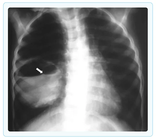
Q23. What is the structure indicated by the arrowhead in the image provided?
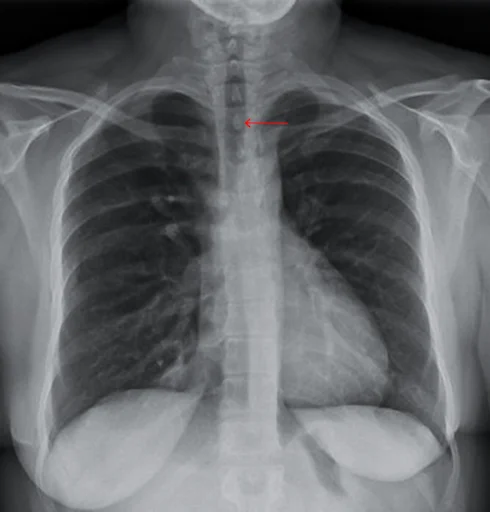
- Esophagus
- Pulmonary artery
- Trachea
- Left atrium
Answer : C - Trachea
Q24. The patient presented with dyspnea and the X-ray indicates:
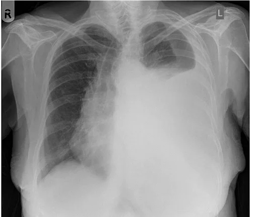
- Consolidation
- Exudative pleural effusion
- Pneumothorax
- Hydropneumothorax
Answer : B - Exudative pleural effusion
Boost your FMGE prep with Test & Explanation—practice real exam questions, learn from detailed solutions, and master concepts with ease.
Download the PrepLadder app now and unlock a 24-hour FREE trial of premium high-yield content. Access Smarter Video Lectures also in हिंglish, Game Changing Qbank, Audio QBank, Structured Notes, Treasures, Mock test for FREE.
Elevate your study experience with PrepLadder’s FMGE online coaching and gear up for success. Start your journey with PrepLadder today!

PrepLadder Medical
Get access to all the essential resources required to ace your medical exam Preparation. Stay updated with the latest news and developments in the medical exam, improve your Medical Exam preparation, and turn your dreams into a reality!