Cardiomyopathy : Types, Causes, Risk Factors, Symptoms and Treatment
Jun 5, 2023

Myocardium (heart muscle) is impacted by the illness known as cardiomyopathy. Cardiomyopathy can develop scar tissue and cause your heart to stiffen, expand, or thicken. Your heart is unable to adequately pump blood to the rest of your body as a result.
Your heart may deteriorate over time, and cardiomyopathy may result in heart failure. Treatment is beneficial. Some cardiomyopathy sufferers eventually require a heart transplant.
Scale up your NEET PG preparation with this blog on important topics for Anatomy and experience the best NEET-PG coaching available online.

Types of Cardiomyopathy
Different forms of cardiomyopathy include:
- Dilated cardiomyopathy: The left ventricle, the heart's primary pumping chamber, enlarges (dilates) in this type of cardiomyopathy and is unable to efficiently pump blood out of the heart. Although this variety can affect people of all ages. Coronary artery disease or a heart attack are the most frequent causes. However, genetic alterations may also be to blame.
- Hypertrophy cardiomyopathy: This type results in aberrant heart muscle thickening, which makes the heart's job more difficult. It mostly affects the left ventricle's (the heart's main pumping chamber) muscle. Although hypertrophic cardiomyopathy can appear at any age, it usually worsens if it does. Although hypertrophic cardiomyopathy can manifest at any age, it usually worsens if it does so in a young child. A family history of the condition is common among those who have this kind of cardiomyopathy. Hypertrophic cardiomyopathy has a hereditary component.
- Restrictive cardiomyopathy: Due to the heart muscle's stiffness and decreased flexibility, this type prevents the heart from expanding and filling with blood in between heartbeats. Although it can afflict anyone at any age, the most typical demographic for this least common type of cardiomyopathy is the elderly.
- Restrictive cardiomyopathy can be the result of a heart illness that affects other parts of the body, such as amyloidosis, or it can develop for no apparent reason (idiopathic).
- Right ventricular dysplasia that is arrhythmogenic. Genetic alterations are frequently the cause.
- Cardiomyopathy that is unclassified: This includes many forms of cardiomyopathy.
Restrictive Cardiomyopathy
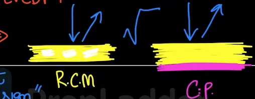
It is the Rarest form of cardiomyopathy. In this we find a condition which is known as STIFF HEART. In this condition there is normal size ventricle but atria is dilated to work against stiff ventricles. Pink Hyaline deposits (amyloid protein) in the myocardium occurs causing the stiffness of the ventricles which is shown in the below image

Causes of Restrictive Cardiomyopathy
- Amyloidosis
- Radiation
- Sarcoidosis
- Hemochromatosis
- Storage disorders: Gaucher disease and Fabry disease
- Scleroderma
- Endocardial fibroelastosis
Clinical Features
- Decreased Right ventricular compliance
- Pitting pedal edema/Ankle edema
- Ascites
- Hepatomegaly/ RUQ (Right upper quadrant) discomfort
- Kussmaul sign: Jugular venous pressure rises with Inspiration
- Decreased Left ventricular compliance
- Pulmonary edema due to pooling of blood in the lungs, Dyspnea on exertion, Orthopnea, Paroxysmal nocturnal dyspnea
- Fibrosis triggers clots in Left atria and ventricle leading to increased chances of embolic stroke
- Effort intolerance
Investigations
- ECG- Low voltage leads due to fibrosis in the heart
- Chest x-ray
- To rule out Constrictive pericarditis (forms calcification around the heart)
- To demonstrate Pulmonary edema
- Trans thoracic echocardiography/ Trans esophageal echocardiography
- Thickness of the Left ventricular and right ventricular wall is normal
- Left ventricular end diastolic pressure is increased
- Cardiac catheterization
- The blood hits the ventricular wall, but it bounces of the wall instead of stretching of the wall due to fibrosis of the ventricular wall
- This is called Square root wave sign due to non-compliant ventricular wall
- Gold standard tool: Endomyocardial Biopsy
- Cardiac MRI- preferred imaging modality in Hypertrophic cardiomyopathy
Treatment
- Implantable cardioverter defibrillator- To increase the chances of survival
- Warfarin: given to reduce the risk of cerebral thrombosis
- Diuretics- Lowest possible dose is given due to low blood pressure
- Cardiac transplant
Dilated Cardiomyopathy
- Globular enlarged flabby heart is seen in dilated cardiomyopathy
- The histopathology shows multinucleated myocytes
- Dicrotic pulse is a feature sign of dilated cardiomyopathy
Causes of dilated cardiomyopathy:
- Sequelae to Viral myocarditis
- Parvovirus B19
- HHV6
- Coxsackie B
- Covid-19
- Toxins- Alcohol
- Sarcoidosis
- Duchenne’s muscular dystrophy
- Functional mitral regurgitation, tricuspid regurgitation- due to the annulus dilatation of the cusp of mitral and tricuspid valves

Investigations
- Trans thoracic echocardiography
- Cardiac MRI
- Chest x-ray- increased cardio thoracic ratio and pulmonary edema
Treatment
- Implantable cardioverter defibrillator
- Cardiac resynchronization therapy
Peripartum Cardiomyopathy
This type of cardiomyopathy mainly takes place from the last month of pregnancy up to 5 months of postpartum. In this we can find conditions such as Pregnancy induced hypertension, valvular diseases. Recovery is possible in peripartum cardiomyopathy.
Takotsubo Cardiomyopathy
This cardiomyopathy occurs due to catecholamine surge. In this case recovery is possible.
Hypertrophic Cardiomyopathy
The heart muscle enlarges as a result of the disorder known as hypertrophic cardiomyopathy (HCM). The thicker heart muscle may make it more difficult for the heart to pump blood. It is typical for hypertrophic cardiomyopathy to be incorrectly diagnosed because many patients have few, if any, symptoms. A small minority of people with HCM, however, may have dyspnea, chest pain, or changes in the electrical activity of the heart, which can result in lethal arrhythmias or unexpected death due to the thicker heart muscle.
Symptoms of Hypertrophic Cardiomyopathy
One or more of the following could be one or more of the signs and symptoms of hypertrophic cardiomyopathy:
- Chest pain, particularly when exercising
- Especially during or right after activity or exertion, fainting
- A healthcare professional might hear a cardiac murmur while listening to the patient's heart.
- Tachycardia
- Breathlessness, especially when exercising
Diagnosis for cardiomyopathy
- ECG
- MRI
- Stress test
Treatment
- Metoprolol (Lopressor, Toprol-XL), propranolol (Inderal, Innopran XL), or atenolol (Tenormin) are examples of beta blockers.
- Verapamil (Verelan, Calan SR) and diltiazem (Cardizem, Tiazac), two calcium channel blockers
- Heart rhythm medications like disopyramide (Norpace) and amiodarone (Pacerone)
- If you have atrial fibrillation or the apical type of hypertrophic cardiomyopathy, which might increase the risk of sudden cardiac death, blood thinners such warfarin (Jantoven), dabigatran (Pradaxa), rivaroxaban (Xarelto), or apixaban (Eliquis) may be prescribed to prevent blood clots and reduce your risk of sudden cardiac death.
Risk Factors For Cardiomyopathy:
Some of the risk factors for cardiomyopathy are beyond your control, such as:
- Your family has a history of heart failure, cardiomyopathy, or sudden cardiac arrest.
- Personal experience with heart attacks.
- Alcohol or cocaine abuse on a long-term basis.
- Pregnancy.
- A situation that causes a lot of stress, such as losing a loved one.
- Chemotherapy or radiation therapy for cancer.
- A BMI (body mass index) more than 30.

Complications of Cardiomyopathy
- A cardiac arrest: The heart cannot physically pump all the blood that the body needs. If untreated, heart failure can be fatal.
- The blood clots: The failure of the heart to pump blood effectively might lead to blood clots developing inside the heart. If clots are put into the bloodstream, they may impede blood flow to other organs like the heart and brain.
- Heart valve problems: Cardiomyopathy can cause the heart to expand, which can prevent the heart valves from shutting properly. As a result, blood may begin to flow backward in the valve.
- Heart arrest and sudden death: Cardiomyopathy can cause abnormal heart rhythms that can make you feel faint or, in rare cases, cause you to pass away suddenly if your heart stops pumping blood adequately.
To scale up your NEET PG preparation with the best-in-class video lectures, QBank, Mock Tests and more, download the PrepLadder App!
Download PrepLadder's NEET PG preparation app for Android
Download PrepLadder's NEET PG preparation app for iOS

PrepLadder Medical
Get access to all the essential resources required to ace your medical exam Preparation. Stay updated with the latest news and developments in the medical exam, improve your Medical Exam preparation, and turn your dreams into a reality!
Navigate Quickly
Types of Cardiomyopathy
Restrictive Cardiomyopathy
Dilated Cardiomyopathy
Causes of dilated cardiomyopathy:
Investigations
Treatment
Peripartum Cardiomyopathy
Takotsubo Cardiomyopathy
Hypertrophic Cardiomyopathy
Risk Factors For Cardiomyopathy:
Complications of Cardiomyopathy
Top searching words
The most popular search terms used by aspirants
- NEET PG Anatomy