Development of the Nervous System - NEET PG Anatomy
Feb 10, 2023

The nervous system development is an important topic for the NEET PG exam because it is crucial for understanding the nervous system's anatomy, physiology and pathophysiology. Understanding the nervous system's development is vital for diagnosing and treating various neurological disorders and diseases.
In the context of the NEET PG exam, knowledge of the development of the nervous system is tested in both theoretical and applied aspects. Questions related to the development of the nervous system can be found in the anatomy, physiology, and neuroanatomy sections of the exam.
The exam also assesses the candidate's ability to apply this knowledge in diagnosing and managing neurological disorders.
Thus, an in-depth understanding of the development of the nervous system is critical for medical students and aspiring doctors for NEET PG Preparation.
Read this blog and get a quick overview of this high-yield topic.

Overview
The nervous system develops from the neural plate ectoderm.
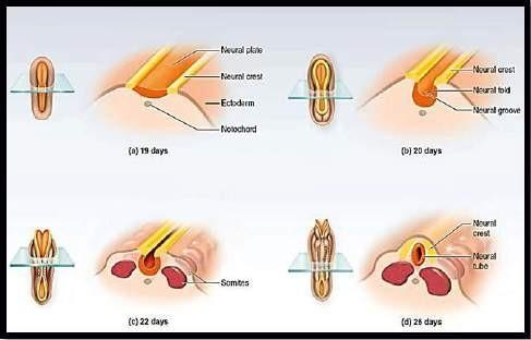
At the dorsum of the baby, a plate develops called the Neural plate. Neural plate develops to form a neural groove, and later neural tube.
Two neural pores are formed:
- Cranial (rostral) neuropore → Closes at day 25, leading to formation of the brain.
- Caudal (posterior) neuropore → Closes at day 28, leading to formation of spinal cord.
This neural plate ectoderm further develops to form a neural groove. This neural groove finally detaches from the dorsal surface and forms the neural tube. This neural tube forms the brain and spinal cord (central nervous system or CNS).
Neural Crest Cells (NCCs):
- The peripheral nervous system (PNS) is formed by the NCCs.
- NCCs are formed at the junction of the neural plate ectoderm and the surface ectoderm.
- NCCs also detach from themselves from the dorsal surface and come to lie dorso-laterally to the neural tube.
- Most of the ganglia are formed by the NCCs.
Important Information:
- Non-closure of anterior neuro-pore Leads to anencephaly
- Non-closure of posterior neuro-pore Leads to Rachischisis
Also Read: Inguinal Canal & Spermatic Cord - NEET PG
OPEN NEURAL TUBE DEFECTS
Open Neural Tube Defects (ONTDs) are congenital anomalies that affect the central nervous system, specifically the brain and spinal cord. They occur when the neural tube, which normally closes during embryonic development to form the brain and spinal cord, does not close properly. This results in an opening in the neural tube, which can cause a range of neurological and developmental problems, depending on the size and location of the defect. Some common ONTDs include

Some common ONTDs include:
- Anencephaly
- It occurs due to non-closure of anterior neuro-pore.
- Brain is small and degenerated without the skull cap.
- Rachischisis
- It occurs due to non-closure of posterior neuro-pore.
- Defect in lumbo-sacral region.
- Vertebra has not fused, spine is bifid.
- Skin is missing over the defect .
- There is leakage of CSF.
- Cranio-rachischisis
- It occurs when there is the total non-closure of the neural tube.
- Therefore, this is a combination of anencephaly and rachischisis.
Also Read: Gametogenesis - Definition, Stages and Types - NEET PG Anatomy
Spina Bifida

Types of Spina Bifida
- Spina bifida occulta
It is the most common type. It is an asymptomatic condition, which is found incidentally and is not evident clinically. Patient might have a tuft of hair in the lumbo-sacral region, but nothing more evident. However, deep down there is a bifid spine. Although, the spine and the meninges are within the limits of the vertebra.
- Spina bifida with Rachischisis
It is the least common type and occurs due to the non-closure of posterior neuro-pore in the lumbo-sacral region. Defect in lumbo-sacral region. Vertebra has not fused, spine is bifid. Skin is missing over the defect and there is leakage of CSF.

- Spina bifida cystica with Meningocele:
A cyst is seen in the lumbo-sacral region which only has protrusion of meninges. Spine is still present within the limits of the vertebra. It is covered by the skin.
- Spina bifida cystica with Meningomyelocele:
A cyst is seen in the lumbo-sacral region with protrusion of meninges and some neural tissue. Spine is outside and lies in the cyst. It is covered by the skin.
Important Information:
- Spina bifida cystica is also known as spina bifida manifesta.
- If the cyst is clear Meningocele
- If the cyst is not clear and has blue/black markings Meningomyelocele (neural tissue present)
Also Read : Development of Skull - NEET PG Anatomy
Fetal Veins, Portal vein formation, Derivatives of the embryonic veins - NEET PG Anatomy

DEVELOPMENT OF VENTRICLES
Ventricles are open spaces within the CNS that contain the CSF. CSF is the ultrafiltrate of blood, secreted by the choroid plexus.
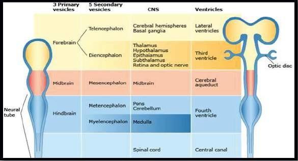
- Neural tube has 3 primary vesicles:
- Prosencephalon (forebrain)
- Mesencephalon (midbrain)
- Rhombencephalon (hindbrain)
- Forebrain develops into:
- Telencephalon
- Diencephalon
- Hindbrain divides into:
- Metencephalon
- Myelencephalon
- Telencephalon divides into:
- Cerebral hemispheres
- Basal ganglia
- Cranial nerve I
- Diencephalon develops into:
- Thalamus
- Hypothalamus
- Subthalamus
- Epithalamus
- Retina
- Cranial nerve II (optic nerve)
- Brainstem is formed by:
- Midbrain
- Pons
- Medulla oblongata
- Cranial nerves III - XII
- Lateral ventricles
- They are ventricles of the cerebrum, one on each side.
- They are the largest of all ventricles, filled with the largest amount of CSF.
- They have most of the choroid plexus (the capillary plexus that forms the CSF).
- Third ventricle
- It is a midline ventricle.
- It is sandwiched between the 2 thalami.
- Cerebral aqueduct of Sylvius
- It is a midline ventricle of the midbrain.
- It connects the third ventricle with the fourth ventricle.
- Fourth ventricle
- It is a midline ventricle of the hindbrain, lying partly in the region of pons and partly in the region of medulla.
- It is a diamond shaped ventricle.
- Pons and upper part of the medulla are present on the floor anteriorly.
- Cerebellum forms the roof of the 4th ventricle posteriorly.
- It continues into the lower medulla as the central canal, which continues further down into the spinal cord.
- Choroid plexus majorly forms the CSF in the lateral ventricle. But it also forms CSF in the third and fourth ventricles.
- CSF escapes from the roof of the fourth ventricle into the subarachnoid space through the following foramen:
- 1 Magendie foramen → In the midline
- 2 Luschka foramen → One on either side of Magendie foramen.
- After the CSF comes into the subarachnoid space, the CSF is absorbed by the dural venous sinuses through the arachnoid villi.

- During the cephalo-caudal folding, the straight neural tube also folds and some part of the brain becomes C-shaped. This also occurs because the brain has to develop in a limited space of the cranial cavity. Hence, the brain bends and becomes spherical.
- Cerebral hemisphere also becomes C-shaped, therefore, the lateral ventricles also become C-shaped.
- Lateral ventricles develop some horns:
- Anterior frontal horn
- Posterior occipital horn
- Inferior temporal horn
- Choroid plexus is found mostly in the lateral ventricles. Though it is also partly present in the third and fourth ventricles. It forms the CSF. The CSF is the ultra-filtrate of blood.
- Flow of CSF:
Lateral ventricles
↓
Third ventricle
↓
Cerebral aqueduct of Sylvius
↓
Fourth ventricle
↓
Central canal
Structures that become C-shaped

Thalamus is the axis around which the various structures are becoming C-shaped. These structures are:
- Cerebrum
- Fornix - Hippocampus sending a C-shaped axons collection
- Choroid plexus
- Caudate nucleus (part of the basal ganglia)
- Corpus callosum
Commissures
- Anterior commissure
It is the 1st commission to develop and connect the right and left side of the brain. For example, the olfactory bulb of the two sides are connected via the anterior commissure.
- Fornix
It is the 2nd commission to develop and is a C-shaped collection of axons. It connects the hippocampus (in the temporal lobe) on either side with each other.
- Corpus callosum
It is the 3rd commissure to develop and connects the right side of the cerebral lobes to the left side of the cerebral lobes.
Hope this blog was insightful. Stay tuned and we’ll keep bringing more such medical notes blog on all the high-yield topics for NEET PG exam to elevate your preparations.
To scale up your NEET PG exam preparation with the best-in-class video lectures, QBank, Mock Tests and more, download the PrepLadder App!
Download PrepLadder's NEET PG app for Android
Download PrepLadder's NEET PG app for iOS

PrepLadder Medical
Get access to all the essential resources required to ace your medical exam Preparation. Stay updated with the latest news and developments in the medical exam, improve your Medical Exam preparation, and turn your dreams into a reality!
Navigate Quickly
Overview
Two neural pores are formed:
Neural Crest Cells (NCCs):
OPEN NEURAL TUBE DEFECTS
Spina Bifida
Types of Spina Bifida
DEVELOPMENT OF VENTRICLES
Structures that become C-shaped
Commissures
Top searching words
The most popular search terms used by aspirants
- Anatomy Important Topics
- NEET PG Anatomy