Regional Injury: Types of Fractures, Intracranial Hemorrhages
May 30, 2023

Regional injuries include wounds to the head, neck, spine, spinal cord, chest, abdomen, and bones and joints of the limbs. These wounds have medical and legal implications. When reconstructing events, the proper interpretation of these injuries is crucial.
Scale up your NEET PG preparation with this blog on important topics for Pathology and experience the best NEET-PG coaching available online.

Head Injury
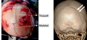
Skull fracture
Most common bone to be fractured is Temporal as it has less thickness.
- Parietal - 5 to 10 mm thickness
- Occipital - 15 mm thickness
- Temporal - 4 mm thickness
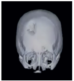
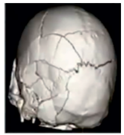
Classification of Skull Fractures:
1. Fissured Fracture
Fissured Fracture is also known as linear fracture. It is the most common fracture of the skull. As it is very thin, 10-15% of these skull fractures are missed even on CT scan. These are obvious during autopsy only.

2. Diastatic fracture
It is associated with sutural separation and is seen primarily in young adults. Most common one is sagittal suture.
3. Depressed Fracture
Depressed fracture is caused by a heavy weapon or object. In this the striking surface of the weapon is small. E.g. Hammer. Small striking surfaces will cause depression and the pattern of fracture tells about the weapon. It is also known as a signature fracture or signature ala fracture. In this the outer surface is destroyed as well as the inner part is destroyed.
4. Pond or Indented Fracture
Generally there is intact dura mater and Inner surface is intact. It is more common in infants. Dent in skull bone is known as pond fracture. It is also known as ping pong ball fracture and is mainly caused by obstetrics forceps blade.
5. Gutter Fracture
This fracture is caused by an oblique bullet which comes tangentially. It is a fracture in the outer surface.This is known as a glancing bullet. Gutter formation in the outer surface.

6. Comminuted Fracture
This fracture occurs by a Heavy blunt blow. In this multiple fragments are formed. Fragments are displaced and there are intersecting lines and webs, known as spider web fracture.
7. Ring or Foramen Fracture
Fracture surrounding foramen magnum. It is seen if someone falls from a height or if falling on feet or falling on buttocks occurs. It Can be due a to blow on the chin a or blow the on vertex
8. Motorcyclist Fracture
It is common in people riding motorcycles. It is known as fracture of the base of the skull. In this force is on the skull that comes from one side to another, mostly in the temporal area. It can be classified in Type 1, Type 2, and Type 3.
Type 1 is a hinge fracture.Type 1 can have a nodding face sign. Type 2 motorcyclist fracture starts from the front area of one side and then to Sella tursica and then to the contralateral side. Type 3 is a fracture in the anterior part of the skull.
9. Bow Out Fracture
This is caused by blunt trauma. It is a fracture of the orbital wall. It can be the median, posterior, or floor of the orbital wall.
Intracranial hemorrhages
First layer is the skull vault or cranial. Then, we have dura mater Arachnoid mater, and Pia mater. Brain tissue/ Cerebrum The space between the skull and dura is almost none. There is an artery known as the middle meningeal artery.
Pterion is a point where the frontal, parietal, sphenoid, and temporal bone meet. This space is known as epidural. The space between the arachnoid and dura is known as subdural space. We have bridging veins that drain to the Dural venous sinus and subarachnoid space. The space between the arachnoid and pia is subarachnoid. Here we have cerebral spinal fluid and many arteries of the circle of Willis.
Also watch a related video on Intracranial Hemorrhage By Dr. Deepak Marwah:
Epidural Hemorrhage
It lies between skull vault & Dura mater.
Patho-physiology:
Any blow/trauma to the side of the head, particularly at pterion results in fracture of temporal bone, leading to the rupture of Middle meningeal vessels.This causes bleeding into extradural space. As this hematoma extends, it causes brain compression & the pt. may die due to respiratory failure.
This is known as EDH. It is like an EDALY shape. EDH is exclusively by trauma. It is on the temporal site. The main part is pterion. EDH is not in the contrecoup region. Minimum amount is 100 ml which can cause compression of the brain and respiratory failure leading to death. Lucid interval is common here.
Lucid Interval
If patient is unconscious and turns conscious and goes unconscious, it is known as a lucid interval. If you commit a crime in this period you will be responsible and In this lucid interval, you can act as a witness or write down a will.
If the patient of head injury having EDH visits the doctor and the doctor does not do a medical examination or CT scan and let the patient go and if the patient then dies then it is considered as medical negligence. It comes under IPC 304A. In this the Punishment could be 2 years and fine.
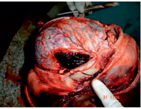
This is Epidural Hematoma. Treatment includes aspiration, burr and hole to remove the blood and Craniotomy
.jpg)
EDH vs Heat Hematoma
EDH HEAT HEMATOMA Caused by trauma 1. Caused by heat/burn Always unilateral 2. It can be bilateral Reddish color 3. Chocolate brown Due to rupture of the middle meningeal artery 4. Contraction of dura mater. Due to the rupture of the Dural venous sinus, there is blood loss. Honeycomb appearance is seen.
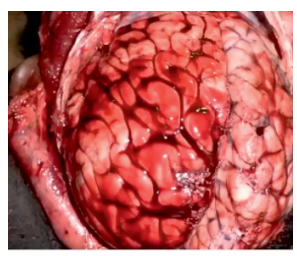
Subdural Hematoma
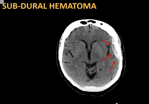
It is sickle-shaped or banana-shaped hematoma. It is concavo-convex shaped. EDH on the brain side is convex and It is due to the rupture of bridging vein SDH is below the dura. It is most commonly caused by trauma. If there is about 100 to 150 ml of blood then it is fatal. Even minor trauma can lead to this.
- Under what condition is this seen?
- Non-traumatic reason is alcohol intake.
- Widespread boxing injury
- Common in children
- Common in elderly people
- It is Sickle shape – concave-convex
- Shaking baby syndrome, which is part of a battered baby syndrome is associated with subdural hematoma.
- SDH can be of 3 types.
- Acute SDH - it occurs in 0 to 3 days and Can be hyperdense.
- Subacute SDH - it occurs in 3 days to 3 weeks and it can be iso-dense.
- Chronic SDH - it occurs in More than 3 weeks - can be hypodense.
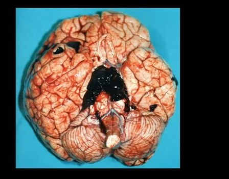
Subarachnoid (SAH)
It is below the arachnoid. SAH can be due to trauma/non-trauma but is most commonly due to trauma. Non-trauma is caused due to rupture of a berry aneurysm. Trauma is caused due to rupture of an artery of Willis.
Most common symptom is a headache which is severe, excruciating pain. This is known as a thunderclap headache. It can lead to nausea and vomiting, sudden loss of consciousness, and neurological problems/photophobia
Diagnosis
In NCCT, the blood will be in the subarachnoid space. Lumbar puncture can show blood in CSF, This is known as Xanthochromia. It is seen in 4 to 6 hours. It is yellow in color due to bilirubin. For SAH, treatment will be endovascular coiling or clipping. To differentiate between SDH and SAH a water test is done. In the test, water is poured and if the blood gets washed off, this is SDH and if the blood is not washed off, it is SAH.
Punch Drunk Syndrome
It is also known as Dementia Pugilistica. It is commonly seen in boxing injuries. It becomes chronic SDH. in this Short-term memory loss is noticed and there isdDisorientation to time, place, and person.
Depression and mood changes can also be seen. Parkinson's symptoms like tremors, and stiffening of muscles can be seen in this.
On autopsy - cerebral atrophy and neurofibrillary tangles can be seen. It is also known as chronic traumatic encephalopathy.
Intracerebral Hemorrhage
It is due to a rupture of the middle cerebral artery. Most common cause is increased blood pressure. The most common site of intracerebral hematoma is the putamen. This is the most common non-traumatic hemorrhage.
- Berlin's edema is seen in concussion injury.
- Commotio cerebri-s - shearing stress in the brain leading to numerous small, punctate hemorrhages throughout the brain.
- Diffuse axonal injury - axonal bulb/retraction bulb (visible after 12 hours) - transacted axon
- Duret hemorrhage - hemorrhage of midbrain and pons.
- Kernohan’s notch - result of raised intracranial pressure.
Frequently Asked Questions
Q: What type of hematoma is convex/ lemon shaped ?
Answer: Epidural hematoma
Q: What type of Hematoma is concave/creasent shaped?
Answer: Subdural Hematoma
Q: Which type of hematoma has a lucid interval?
Answer: Epidural Hematoma
Q: Which bone is the most prone to skull fractures?
Answer: Temporal bone is most prone to fractures as it is the least thick.
Q: What is the most common cause of EDH and SDH?
Answer: Head Injury
Q: What is the weakest part of the skull?
Answer: Pterion
If you're looking to test your preparation and gear up for the NEET PG exam, the NEET PG Mock exam is always available for you. This comprehensive mock exam, designed to mirror the actual exam pattern and difficulty, is available throughout your preparation to assess your progress.
To scale up your NEET PG preparation with the best-in-class video lectures, QBank, Mock Tests and more, download the PrepLadder App!
Download PrepLadder's NEET PG preparation app for Android
Download PrepLadder's NEET PG preparation app for iOS

PrepLadder Medical
Get access to all the essential resources required to ace your medical exam Preparation. Stay updated with the latest news and developments in the medical exam, improve your Medical Exam preparation, and turn your dreams into a reality!
Navigate Quickly
Head Injury
Skull fracture
Classification of Skull Fractures:
Intracranial hemorrhages
Epidural Hemorrhage
Lucid Interval
EDH vs Heat Hematoma
Subdural Hematoma
Subarachnoid (SAH)
Diagnosis
Punch Drunk Syndrome
Intracerebral Hemorrhage
Frequently Asked Questions
Q: What type of hematoma is convex/ lemon shaped ?
Q: What type of Hematoma is concave/creasent shaped?
Q: Which type of hematoma has a lucid interval?
Q: Which bone is the most prone to skull fractures?
Q: What is the most common cause of EDH and SDH?
Q: What is the weakest part of the skull?
Top searching words
The most popular search terms used by aspirants
- NEET PG Strategy