Pathology of Brain Tumors: Types, Grading, and Diagnosis
Nov 19, 2024

A brain tumor is a peculiar growth of cells within the brain or central spinal cord. These tumors can be both primary (originating in the brain) or secondary (metastatic, originating somewhere else and spreading to the brain). Brain tumors can vary broadly in terms of their kind, location, and behavior, ranging from benign (non-cancerous) to malignant (cancerous).
Primary Brain Tumors: Grading
A - Atypia
M - Mitosis
E - Endothelial cell proliferation
N - Necrosis
- Grade 1: Atypia, mitosis, endothelial cell proliferation, and necrosis are absent.
- Grade 2: Atypia is present.
- Grade 3: Atypia and mitosis are present.
- Grade 4: 3/4 features are present.
Classification of Brain Tumors
Classification of Brain Tumors
Brain Tumors Types Gliomas: glial cells - Astrocytoma
- Oligodendroglioma
- EpendymomaNeuronal tumors - Ganglioglioma: most common
- Gangliocytoma
- Neurocytoma
- DNET (Dysembryoplastic neuroepithelial tumor)
- Floating neurons in pools of mucopolysaccharideUndifferentiated tumors - Medulloblastoma
- Atypical teratoid/atypical rhabdoid
- SNF/INI mutation - rhabdoid tumor shows SNF mutantMeningeal tumors
Gliomas: glial cells
Astrocytoma
| Grade 1 | - Juvenile Pilocytic Astrocytoma (JPA): Small tumor - Subependymal Giant Cell Astrocytoma (associated with tuberous sclerosis) |
| Grade 2 | - PXA - Diffuse astrocytoma - Fibrillary astrocytoma |
| Grade 3 | Anaplastic astrocytoma |
| Grade 4 | Glioblastoma |
Also read: Important Pathology Questions on Cell Injury – MCQs & Key Concepts
Juvenile Pilocytic Astrocytoma: CCC factor
| Age | Children |
| Site | Cerebellum |
| Gross | Cystic lesion with a mural nodule |
| Mutation | BRAF-KIAA fusion BRAF V E mutation |
| Microscopy | - Rosenthal fibers (thread-like structure, non-specific) - Mulberry body - Composition: alpha-beta crystallin, ubiquitin, heat shock proteins, and GFAP. 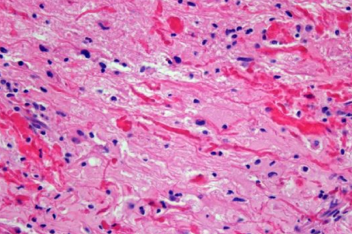 |
| Prognosis | Good |
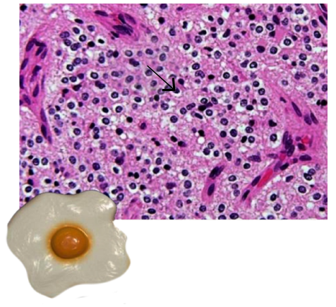
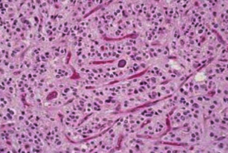
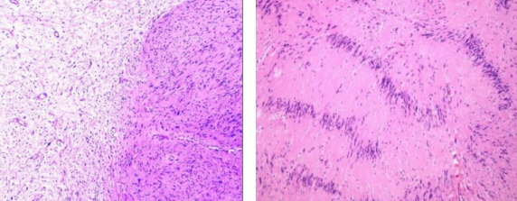
Glioblastoma
| Age | Most common malignant brain tumor in adults |
| Gross | Resembles a butterfly - crosses the midline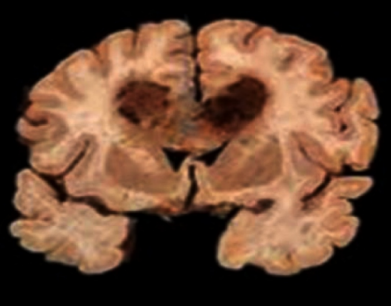 |
| Microscopy | Serpentine necrosis - surrounded by pseudo-palisading of the tumor cells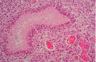 Glomeruloid Body - Endothelial cell proliferation 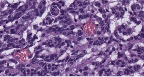 |
| Mutation | IDH - wild type (90%) - p53 - PTEN - EGFR - TERT IDH - mutant type ( problem) - p53 - ATRX - TERT |
| Prognosis | Very bad |
Also read: Plasma Cell Dyscrasia & Flow Cytometry in Hematology
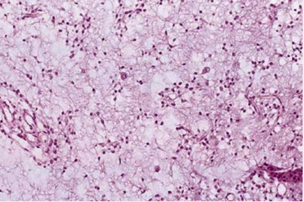
Oligodendroglioma
- Site - Frontal Lobe
- Gross - Calcification +
- C - Craniopharyngioma
- O - Oligodendroglioma
- M - Meningioma
- Microscopy -
- Fried egg appearance: nucleus and & Perinuclear hal
- Chicken wire appearance of blood vessel
- Perineuronal satellitosis
- Mutation -
- IDH mutation: most common
- Co-deletion 1p/19q-good response to chemotherapy
| Fried egg appearance | Chicken wire appearance |
- Brain-oligodendroglioma - Bone marrow biopsy-hairy cell leukemia - Testes/Ovary-seminoma/ dysgerminoma - Microbiology colonies - Malassezia furfur - Mycoplasma | - Blood vessels - Oligodendroglioma - Breast FNAC (mucinous/colloid carcinoma) or Fibrosis: ALD o Calcification: chondroblastoma |
Also read: Comprehensive Overview Of Lung Pathology Images
Ependymoma
Grade II tumor
| Site | Children, 4th Ventricle Adults: Spinal Cord |
| Genetics | MIS M E - Multiple Inherited - Schwannoma - Meningiomas - Ependymoma - Neurofibromatosis-2 |
| Microscopy | Perivascular Pseudorosettes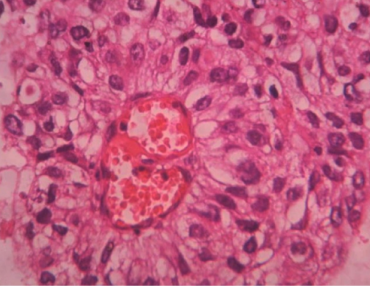 |
Updates
Myxopapillary Ependymoma
- Myxoid + Papillae
- Located in Filum Terminale
- Grade 1 tumor
Rela Fusion Ependymoma
- R - Rela gene
- EL - chr 11
- A - above tentorium in the brain/supratentorial tumor
- Occurs in children
- Poor prognosis
Neuronal Tumors
- Ganglioglioma: most common
- Gangliocytoma
- Neurocytoma
- DNET (Dysembryoplastic Neuroepithelial Tumor)
- Floating neurons in pools of mucopolysaccharide
Also read: Blood Groups and Storage in Blood Banking
Undifferentiated Tumors
Medulloblastoma - Grade IV tumor
| Age | Child |
| Site | Cerebellum (not cystic) |
| Drop Mets | Cerebellum →Spinal cord (CSF) |
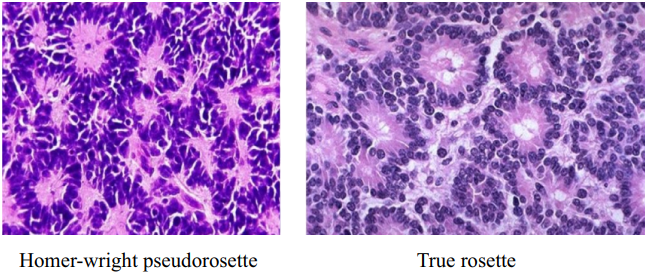
| Microscopy | Homer Wright pseudorosette (non-specific) M - Medulloblastoma E- Ewing's sarcoma N– Neuroblastoma |
- Pseudorosettes: presence of material/blood vessels in the center.
- Homer Wright pseudorosette (M/E/N): pink material in the center
- Perivascular pseudorosette—blood vessel in the center (seen in ependymoma)
- True rosette: absence of material/blood vessel in the center (appears clear/empty).
- Flexner-Wintersteiner rosette seen in retinoblastoma.
Medulloblastoma: Genetics
| 1. WNT pathway | Best prognosis |
| 2. SHH pathway N-myc amplification | Intermediate prognosis |
| 3. Non-WNT non-SHH pathway N-myc amplification and i17q | Worst prognosis |
| 4. Non-WNT, non-SHH pathway i17q | Intermediate prognosis |
Also read: Acute Lymphoblastic Leukemia: Symptoms, Causes and Treatments
Gorlin syndrome
- PTCH gene mutation
- SHH pathway is affected
- Desmoplastic Medulloblastoma
- Basal cell carcinoma
- Bilateral ovarian fibroma
Note : TURCOT syndrome
- Colon tumor (COT)
- Brain tumor : medulloblastoma (TUR - turban)
Atypical Teratoid/Atypical Rhabdoid- AT/AR
- SNF/INI Mutation
- Atypical rhabdoid : shows SNF mutation
Meningioma
- Risk factors
- Females
- Pregnancy
- Radiation
- Nf2
- Progesterone receptor +ve
- It is a dura-based tumor—whorling of the tumor cells
- Psammoma bodies
- Grade depends on the progression of tumor
Also read: NEET PG High Yield Questions for Pathology
Miscellaneous
Lymphoma of brain
- EBV infection in HIV-positive individuals
- Most common is DLBCL
- Angiocentric pattern
- Hooping pattern
Schwannoma
- Nf2
- It can occur at any site.
- Spinal cord: dumbbell-shaped appearance
- Microscopically:
- Antony A: hypercellular area
- Antony B: hypocellular area
- Verocay bodies (two rows of palisading nuclei with cytoplasm in the center)
- MPNST is different from schwannoma so mention it as different heading
Malignant peripheral nerve sheath tumor (MPNST)
- Malignant version of schwannomma
- Triton tumor: MPNST+ rhabdomyoblasts
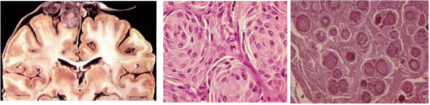
Craniopharyngioma
- Origin: Rathke's pouch
- Site: Suprasellar location + calcification
- Age: Children
- Clinical feature: vision disturbance & headaches
- Two types : Adamantinomatous and Papillary
- Palisading nuclei
- Stellate reticulum
- Wet keratin on gross examination : machine-oil appearance
Update: Diffuse Midline Glioma
- Occurs in the midline of the brain
- Seen in children
- Glial tumor
- It spreads out
- Bad prognosis
- H K M-mutation (H3 is histone)
Also read: Renal Cell Carcinoma
Important questions
Q. Which is the most common malignancy in the brain/lungs/heart/liver/bones?
Ans. Metastasis
Q. Most common cancer to cause brain metastasis?
Ans. Small cell lung cancer
Q. Cancer that never metastasizes to the brain?
Ans. Prostate cancer
Q. Which is the cancer that metastasizes to the meningeal part?
Ans. Leptomeningeal carcinomatosis commonly from breast cancer
.jpg)
MCQs
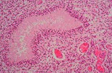
Q. A 10-year-old male child shows a well-circumscribed cystic lesion in the cerebellum. A mural nodule is identified within the cystic lesion. Biopsy and microscopic evaluation show the following features: What is your diagnosis?
- Oligodendroglioma
- Glioblastoma
- Meningioma
- JPA
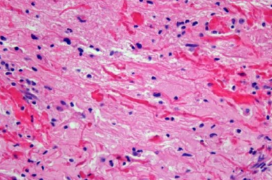
Q. A 55-year-old man presents with seizures and muscle weakness but no other neurological signs. A CT scan reveals a mass in the left cerebral hemisphere. A left frontotemporal craniotomy is performed. A histological examination of the brain biopsy is shown.
- Oligodendroglioma
- Glioblastoma
- Meningioma
- JPA
Also read: Trinucleotide Repeat Disorder (Fragile X Syndrome, Huntington's disease)
Q. A 35-year-old female patient presents with epileptic attacks. Radiological investigations reveal the presence of a frontal lobe mass with foci of calcification. Biopsy shows the presence of sheets of cells with a perinuclear halo. The patient is diagnosed with a Grade 2 brain tumor and advised genetic analysis. Treatment of the patient shows a good response to chemotherapy. Which of the following tumors best describes this?
- Oligodendroglioma
- Glioblastoma
- Meningioma
- JPA
Q. A 33-year-old female with acoustic schwannoma. She also shows the presence of pigmented lesions over the trunk and lower back. On radiological investigations, she has a non-infiltrating brain tumor showing areas of calcification. On biopsy, the tumor shows positivity for PR. Diagnosis.
- Oligodendroglioma
- Glioblastoma
- Meningioma
- JPA
Download the PrepLadder app now to access high-yield content with 24-hr Free Trial. Explore premium study resources like Video Lectures, digital notes, QBank, and Mock Tests for a seamless exam preparation. Time to begin your NEET PG coaching online with PrepLadder.

PrepLadder Medical
Get access to all the essential resources required to ace your medical exam Preparation. Stay updated with the latest news and developments in the medical exam, improve your Medical Exam preparation, and turn your dreams into a reality!
Navigate Quickly
Primary Brain Tumors: Grading
Classification of Brain Tumors
Classification of Brain Tumors
Gliomas: glial cells
Neuronal Tumors
Undifferentiated Tumors
Meningioma
Miscellaneous
Important questions
MCQs
Top searching words
The most popular search terms used by aspirants
- NEET PG Pathology
- NEET PG Pathology Preparation
PrepLadder Version X for NEET PG
Avail 24-Hr Free Trial