Breast Cancer - Causes, Diagnosis and Treatment
May 10, 2023

Breast cancer comes in a variety of forms, including: invasive (infiltrating) ductal carcinoma. This cancer starts in your breast's milk ducts, breaks through the duct wall, and then spreads to the adjacent breast tissue. This is the most prevalent kind of breast cancer, accounting for around 80% of all occurrences. Men and women can both develop breast cancer, although women are much more likely to do so. Breast cancer diagnosis and treatment have evolved as a result of significant expenditure in research and awareness campaigns. Thanks to earlier detection, a revolutionary tailored therapy approach, and a better knowledge of the disease, breast cancer survival rates have grown and the number of fatalities linked to the disease is rapidly reducing.
After skin cancer, breast cancer is the second most frequent malignancy among women. The majority of those affected are anticipated to be women over 50.Breast cancer can affect men, however it is uncommon. Male breast cancer affects about 2,600 males annually in the US, accounting for fewer than 1% of total cases.
Breast cancer is more common in transgender women than cisgender men. In comparison to cisgender women, transgender men had a lower risk of developing breast cancer.
Read this blog further to get a quick overview of this important topic for pathology and ace your NEET PG exam preparation.

Types of breast cancer
There are numerous types of breast cancer, including:
- invasive ductal carcinoma (infiltrating). This cancer starts in your breast's milk ducts, breaks through the duct wall, and then spreads to the adjacent breast tissue. With over 80% of cases being this type, it is the most common type of breast cancer.
- ductal carcinoma in situ. Some people consider ductal carcinoma in situ, or Stage 0 breast cancer, to be precancerous because the cells haven't spread past your milk ducts. This condition is remarkably treatable.
- invasive lobular cancer, which has infiltrated. This cancer grows in the lobules of your breast, which are where breast milk is generated. The importance of frequent mammography and clinical breast exams for women with lobular carcinoma in situ is clear.
- Triple negative breast cancer is referred to as TNBC. One of the most difficult breast cancers to treat is triple negative breast cancer, which makes up 15% of cases overall. It is referred to as triple negative breast cancer because it lacks three of the markers connected to other kinds of breast cancer. This makes diagnosis and therapy challenging.
- Inflammatory breast cancer.
Classification of Breast Tumors
- Breast tumors are Classified as two main classes:
- Epithelial
- Stromal
The two kinds of epithelial breast tumor are:
- Ductal-Ductal is categorized as a precancerous type of ductal carcinoma in situ (DCIS). Ductal cancer with infiltration (IDC).
- Lobular- Lobular is categorized as a precancerous type of lobular cancer in situ (LCIS). ILC, invasive lobular cancer.
Stromal Breast Tumor have 2 subtypes:
- Intralobular tumors
- Interlobular tumors
Intralobular tumors: Rises from the lobule.
- Fibroadenoma
- Phyllodes Tumor
Interlobular tumors
- Lipoma
- Angiosarcoma
- Myofibroblastoma
Stromal tumors:
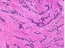
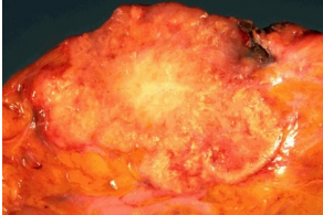
Fibroadenoma- A solid, not a fluid-filled lump, fibroadenoma is a benign (non-cancerous), unilateral breast tumor that causes no pain. Although it can happen at any age, it most frequently affects females between the ages of 14 and 35. In postmenopausal women, fibroadenomas are less frequent because they shrink after menopause.
Clinical presentation of fibroadenoma is it is movable, soft to hard lump.
On Radiology examination we can find breast mouse - The lump is movable and slippery, that's why it's called "breast mouse."
It also demonstrates popcorn calcification (researched under pulmonary tumors)
There are two types of popcorn calcification:
- Radiologically Calcification is a feature of pulmonary hamartomas and fibroadenomas.
- Nodular lymphocyte predominant Hodgkin's lymphoma (NLPHL) RS cells exhibit pathological calcification.
Radiographically, pulmonary hamartoma exhibits coin lesion with popcorn calcification.
Changes in the MED12 gene have been linked three times to pathology.
- Fibroadenoma
- Leiomyoma, or Phyllodes Tumor Fibroids
- Mnemonic: MED in the family
Drug connected to bilateral fibroadenomas: Cyclosporine
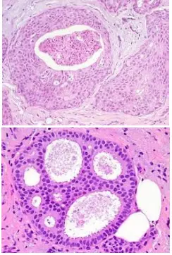
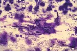
FNAC of fibroadenoma

- When a patient arrives with Breast mouse FNAC is performed. Findings are:
- Staghorn clusters of cells - deer horn like cells.
- Bare nuclei - cytoplasm is stripped off and only nuclei are visible in blue color.
- Other Staghorns
- Staghorn calculus - Present on kidneys called struvite calculus, made of triple phosphatase.
- FNAC - Fibroadenoma
- Blood vessels - Hemangiopericytoma
- Megakaryocyte - Essential Thrombocytopenia.
Histopathological Findings
- Intracanalicular fibroadenoma - In this we will find that the Ducts are compressed.
- Pericanalicular fibroadenoma - In this we will find that the ducts are open
Phyllodes Tumor
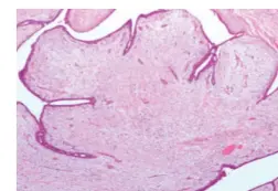
It was Earlier known as cystosarcoma phyllodes. It is a stromal tumor. Phyllodes means phylum which is a leaf-like structure. Earlier it was known that there may be a cyst which may or may not be cancerous. Phyllodes tumors can be benign or malignant.
Epidermal Tumors
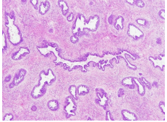
1)Ductal Carcinoma In Situ
- It is a Precancerous situation. The atypical cells fill up the ducts. Both the myoepithelial cells and basement membrane are present. When these cells and membrane vanishes, it can be termed as cancer.
Types of Ductal Carcinoma In Situ(DCIS)
Comedo-necrosis, cribriform DCIS.
It is Solid DCIS-in this the Duct is full of cells. So it Becomes solid. It has following types:
- Papillary: The cells have made small proper papillary.
- Micropapillary: The cells make very tiny papillary.
- Comedo-necrosis: Necrosis in the middle of the duct.
- Just like the dirt that comes out from the acne.
- Cribriform: Meaning sieves
- The duct contains holes within it.
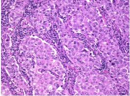
2. Infiltrating Ductal Carcinoma
- It is Infiltrating Ductal Carcinoma, not otherwise specified. It is not a special type of cancer. Gross appearance: Gritty due to calcification, fibrosis. Microscopically it appear as Adenocarcinoma
Special Types of IDC
a)Tubular carcinoma: It shows the presence of regular tubules. This carcinoma Have a good prognosis.
b)Mucinous or Colloid carcinoma:it Has pools of mucin extracellularly and may be intracellularly (called signet ring cell). FNAC: shows chicken wire blood vessels. On Biopsy: it Shows more mucin both extracellular and intracellular with signet rings. Floating cells
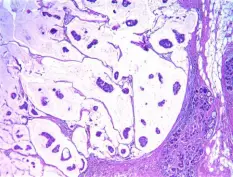
Clinical findings of mucinous carcinoma:
- It is Soft to firm lump due to mucilage. Normally cancerous tumors are hard. So this can be mistaken for a benign tumor.
c) Medullary carcinoma
- Medullary carcinoma presents with:
- Pushing membrane
- Prominent nucleoli
- Lymphoplasmacytic infiltrate - The only visible presentation.
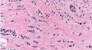
3. Invasive Lobular Carcinoma
The Genetic cause of invasive lobular carcinoma is CDH1 gene mutation. In this the E-cadherin levels decrease. Patient presents with Bilateral or Multicentric nodules.
Microscopy of ILC
- In this the Tumor cells are present back to back, just like the army men.Hence, called Indian File Pattern. It Also shows minimal desmoplasia (less fibrosis) - tumor is not hard. The ducts are unaffected and normal but surrounded by the tumor cells Called targetoid spread. The tumor cells try to enter the normal ducts called the pagetoid spread.
Stages of breast cancer
The staging procedure describes how far advanced the cancer is in your body. This choice is influenced by a number of factors, including the tumor's size, location, and extent, as well as if the disease has spread to other areas of your body. Breast cancer's initial stages are:
- Stage 0. The disease is not intrusive. This shows it hasn't come out of your breast ducts.
- Stage I cancer cells have already migrated into the nearby breast tissue.
- Second stage. The cancer either has a diameter of less than 2 centimeters and has spread to the lymph nodes under the arms, or it has a diameter of more than 5 centimeters but hasn't. Tumors may or may not affect the nearby lymph nodes at this stage, and size ranges between 2 and 5 centimeters.
- Stage three. It might have expanded to neighboring lymph nodes and tissue, but it hasn't reached distant organs. It is referred to as locally progressed.
- Stage four. In this stage the cancer has spread to other organs apart from the breast such as bones, liver, lungs, or brain. Metastatic breast cancer is another name for stage IV breast cancer.
Causes of breast cancer:
- Age-age over 50 years of age increases chances of breast cancer.
- Sex-women are more likely to be diagnosed with breast cancer.
- Family history and genetics- any family history of breast cancer can increase chances of being a victim of breast cancer.
- Smoking
- Alcohol intake .
- Obesity-it can increase chances of having breast cancer and also its recurrence.
- Radiation exposure
- Hormone replacement therapy: women on oral contraceptive pills or other hrt are at higher risk for developing breast cancer.
Also Read:

Risk Factors of Breast Cancer
- Sporadic factors: Non familial
- Age
- Early menarche - increases estrogen levels.
- Late menopause - increases estrogen levels.
- Nulliparity
- Sedentary lifestyle, obesity, high-fat diet.
- Smoking
- Genetic factors: Non Familial
- Related to BRCA1 and BRCA2 - comprises 90% of Familial breast cancer.
- Recalling Neoplasm chapter,
- BRCA1 - present on chromosome 17.
- BRCA2 - present on chromosome 13.
DIAGNOSIS OF BREAST CANCER:
A breast exam will be performed by your healthcare provider, who will also inquire about your family history, medical history, and any current symptoms. In order to screen for breast abnormalities, your doctor may also advise certain tests. These examinations could involve
Mammogram. Your breast alterations or abnormal growths can be seen on these specialized X-ray images. In order to prevent breast cancer, mammography is frequently used.
Ultrasonography. To photograph the tissues inside your breast during this test, sound waves are used. Breast lumps or other abnormalities can be diagnosed with its assistance.
Positron emission tomography (PET) imaging Unusual spots are highlighted during a PET scan using specific dyes. A specific dye is injected into your veins during this test, and the scanner is used to capture photos.
MRI stands for magnetic resonance imaging, and it is a procedure that creates crystal-clear, finely detailed images of the internal breast structures using magnets and radio waves.
They might do a breast tissue biopsy if your doctor notices anything worrisome on the imaging tests. A pathology lab will be contacted to do the analysis on the material.
TREATMENT OF BREAST CANCER:
many treatments available such as surgery, chemotherapy, radiation ,hormone therapy, immunotherapy, targeted drug therapy. What's best for you will depend on a number of factors, including the size and location of the tumor, the results of your lab tests, and whether the cancer has spread to other parts of your body for breast cancer surgery
During breast cancer surgery, the affected portion of your breast as well as the area of healthy tissue surrounding the tumor are removed. There are several types of surgery, depending on your circumstances, including:
- Lumpectomy. A lumpectomy, also referred to as a partial mastectomy, entails the excision of the tumor along with a small margin of surrounding healthy tissue. The majority of the time, a few lymph nodes in the breast or under the arm are also taken out for examination. Radiation therapy is routinely given to the patient in the days and weeks after a lumpectomy.
- Mastectomy. You also have the option of having your entire breast removed. In certain situations, a nipple-sparing mastectomy may maintain your nipple and areola (the black skin around your nipple). Many women choose to have either immediate or delayed breast reconstruction after a mastectomy.
- A common option is breast reconstruction.
- a sentinel node biopsy. Because early-stage breast cancer often results in the lymph nodes being negative (for cancer), the sentinel node biopsy was developed to prevent the wasteful removal of a considerable number of lymph nodes that were not affected by the malignancy. To find the sentinel lymph node, doctors inject a dye that tracks to the lymph node where cancer would first spread. If a lymph node is cancer-free, further lymph nodes do not need to be removed. If one lymph node has cancer, additional lymph nodes might also need to be removed. Even though several sentinel nodes are usually discovered, the chance of developing arm swelling (lymphedema) lowers the fewer lymph nodes that are removed.
OTHER treatment for breast cancer which are available are:
Chemotherapy:
Your doctor can suggest breast cancer chemotherapy before conducting a lumpectomy in an effort to shrink the tumor. It is occasionally given after surgery to get rid of any cancer cells that could still be there and lessen the chance of a recurrence. If the cancer has moved outside of your breast to other parts of your body, your doctor may advise chemotherapy as your main course of treatment.
Radiation therapy for breast cancer:
Radiation therapy for breast cancer is routinely used to eliminate any remaining cancer cells following a mastectomy or lumpectomy. Furthermore, it can be utilized to treat particular metastatic tumors that are bothersome or causing other problems.
Hormone therapy:
Some breast cancer types are assisted in growing by hormones like progesterone and estrogen. Hormone treatment is most typically used by medical experts after surgery to reduce the likelihood of breast cancer recurrence. However, another option is to treat cancer that has spread to other bodily areas or to shrink the tumor before surgery.
Prevention:
Although breast cancer cannot be entirely avoided, there are a number of things you can do to reduce the likelihood that you will discover it when it is already somewhat advanced. For illustration:
Get routine mammograms. The American Cancer Society suggests beginning mammograms for screening at age 35 and continuing them every year beyond age 40.
Once you are 20, you should monthly inspect your breasts. You'll grow acclimated to the size, texture, and form of your breasts and become more alert to changes as a result.
Have your breasts examined by a medical practitioner after the age of 20, and each year beyond the age of 40. Clinical breast exams can detect lipos that mammograms might miss.
This is everything that you need to know about hyperthyroidism for your pathology preparation. For more interesting and informative blog posts like this download the PrepLadder App and keep reading our blog!

PrepLadder Medical
Get access to all the essential resources required to ace your medical exam Preparation. Stay updated with the latest news and developments in the medical exam, improve your Medical Exam preparation, and turn your dreams into a reality!
Navigate Quickly
Types of breast cancer
Classification of Breast Tumors
The two kinds of epithelial breast tumor are:
Stromal Breast Tumor have 2 subtypes:
Stages of breast cancer
Causes of breast cancer:
Risk Factors of Breast Cancer
DIAGNOSIS OF BREAST CANCER:
TREATMENT OF BREAST CANCER:
OTHER treatment for breast cancer which are available are:
Prevention:
Top searching words
The most popular search terms used by aspirants
- Medical PG Pathology
- NEET PG Pathology