Neural Regulation of Respiration: Control & Mechanisms
Nov 7, 2024

Voluntary Control of Respiration
- Voluntary control means breathing can be controlled according to one's wish.
- People can voluntarily hold their breath. However, holding the heart is impossible because the cortex, controlling the regulatory center, has no direct control over the heart.
- Voluntary control of respiration is done by the cerebral cortex, which influences the spinal cord and respiratory centers.
Grab the Most Important 100 One - Liners in Physiology - Download Free PDF!
Automatic Control of Respiration
Two important areas of the central nervous system are responsible for the automatic control of respiration
1. Pons
- The upper part of the Pons contains the pneumotaxic center and
- The lower part of the pons consists of the apneustic center.
- Both are responsible for controlling the rate and the depth of breathing.
- The pneumotaxic center mainly controls the rate of breathing.
- The apneustic center mainly controls the depth of breathing.
2. Medulla
The pacemaker, the start for respiration, or the place where the respiratory rhythm originates is the medulla. The medulla consists of the following:
The dorsal respiratory group of neurons (DRG)
- Includes a collection of neurons close to the nucleus tractus solitarius (NTS, center for various afferents from various viscera).
- DRG is the sensory afferent for the respiratory center.
- DRG integrates sensory signals from the peripherals, such as the lungs or the muscles.
- This is the main function of DRG, and it is not responsible for respiratory rhythm generation.
The ventral respiratory group of neurons (VRG)
- Divided into four parts, among which one of the parts responsibilities is pace-making activity of respiration.

- As seen above, the pneumotaxic center is close to the Kölliker-Fuse and parabrachial complex.
- Just below the pneumotaxic center, the apneic center is present.
- Close to the nucleus tractus solitarius, the collection of the neuronal group is the dorsal respiratory group (DRG).
- The large group of neurons, as can be seen highlighted in green in the diagram, is the ventral respiratory group of neurons (VRG).
- The ventral respiratory group of neurons consists of four different clear zones.
- Bötzinger complex (BötC).
- Pre-Bötzinger complex.
- Then, the rostral ventral respiratory group neurons and
- The caudal ventral respiratory group neurons can be seen.
- Professor Jack L. Feldman first discovered the Bötzinger complex, which plays a very important role in controlling respiration. The name Bötzinger comes from a famous white wine.
- Feldman and colleagues later discovered that the Bötzinger complex is not an important center for respiration, but a center located behind, known as pre-Bötzinger, is important.
- Thus, the pre-Bötzinger complex is considered the main center for respiration and is known as the pacemaker of respiration.
Also read: Hyponatremia: Classification, Signs & Symptoms
Ondine's Curse
- This condition gets its name from German mythology.
- Ondine was a beautiful water nymph who fell in love with a man named Hans. Hans promises he will not marry a second woman ever but later turns unfaithful.
- When Ondine learns that Hans married a second woman, she angrily curses Hans. The curse stated that Hans could not sleep all his life.
- Ondine wants to say that Hans will have no automatic control of respiration. The voluntary control of respiration, in this case, will remain intact.
- With no automatic control of respiration, the pontine and medullary centers are damaged.
- But the cortical control stays intact.
- Cortical control means that when one is awake, the cortex is awake, and one can control their breathing.
- When one goes to sleep, the cortical control is lost as the cortex also goes to sleep, and the respiration will stop.
- When respiration stops, the CO accumulates in the body, causing dyspnea.
- The person will then wake up. Again, when they feel sleepy and fall asleep, they will wake up.
- Hans would thus not be able to get complete sleep.
- This will continue until there is severe exertion of all respiratory muscles, eventually leading to death.
Also read: Hypernatremia: Causes, Pathophysiology
Clinical Versions of the Ondine's Curse
- There are certain congenital disorders, such as congenital central hypoventilation syndrome.
- This is equivalent to Ondine's Curse.
- In the case of bulbar poliomyelitis, when polio affects the pontine and medullary centers, these are destroyed, automatic control is lost, and voluntary control is intact.
- Certain tumors in the medullary center can also produce this kind of clinical picture.
- After an injury, this kind of clinical picture can also be seen.
- In these cases, the only support that can be given is ventilatory support.
.jpg)
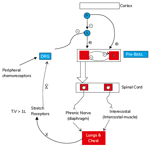
How are the Neuronal Centres Connected?
Pre-Bötzinger Functions
- Acts as the respiratory pacemaker for spontaneous inspiration and expiration.
- Contains I (inspiration) and E (expiration) neurons with spontaneous discharging properties.
- These neurons are spontaneously discharged due to their membrane properties and certain channels on their membranes.
- But the particular property by which it can produce spontaneous inspiration and expiration is unknown.
- For example, in the cardiac system, the mechanism of SA nodal pace-making activity is known, and why it is generating spontaneous rhythm.
- Here, since there is spontaneous discharge of neurons, it is known as the pacemaker neuron. This pacemaker property is due to certain membrane channels, which are unknown.

Also read: NEET PG High Yield Questions for Physiology
Neuronal Discharge Process
- The neuron will start discharging and remain active for 2-3 seconds.
- When the I neuron discharges, it will have an inhibitory signal on the E neuron.
- After 2-3 seconds, the E neuron is activated automatically, which will suppress the I neuron when activated.
- There will then be a pattern of inspiration followed by expiration.
Axonal Extension
- The I neuron is extended up to the level of the spinal cord.
- This means that the axons of this neuron are projected at the spinal cord's level, where they influence spinal neurons.
- Here, the cervical and thoracic regions of the spinal cord are involved.
- The I neuron impulses the phrenic nerve (cervical) and the intercostal nerve (thoracic).
Respiratory Muscle Activation
- Because of this, when the I neuron is activated, it activates the phrenic nerve (cervical) and the intercostal nerve (thoracic) at the level of the spinal cord.
- This causes the diaphragm and intercostal muscles to contract, which causes expansion of the lung and chest.
- When the I neuron discharges, it inhibits the E neuron.
- After 2-3 seconds, when the E neuron is activated, it will inhibit the I neuron. This will cause the diaphragm and intercostal muscles to be relaxed. This is expiration.
- Expiration is thus passive because there is no direct influence on the spinal cord. Inhibition of the I neuron is sufficient to cause expiration.
Voluntary Control and Regulation
- The cerebral cortex controls the pre-Bötzinger complex neuron for voluntary respiration.
- The pneumotaxic center and apneustic center are also responsible for controlling the pre-Bötzinger.
- The pneumotaxic center is seen in the upper part of the pons, and the apneustic center is in the lower part of the pons.
- The apneustic center (A) is a tonically active center that is always active until other neurons inhibit it.
- The apneustic center, or A neurons, are always discharging. There are two inhibitors of this:
- The pneumotaxic center has inhibitory control.
- The vagus nerve.
Also read: NEET PG Previous Year Question Papers of Last 6 Years
Stretch Receptor Activation
- Stretch receptors in lung smooth muscles activated during lung and chest expansion.
- Signal sent to the dorsal respiratory group of neurons, acting as the sensory integration center.
- From the periphery signal from stretch receptors in bronchi and bronchioles transmitted via the vagus neuron.
Apneustic center role
- From the dorsal respiratory group of neurons, the inhibitory input goes to the apneustic center.
- It is to be noted that when the tidal volume is greater than 1 liter (Normal volume- 500 ml), the stretch receptors of the lungs will be activated.
- The apneustic center has a strong stimulatory control over the Inspiratory (I) neuron, which controls the depth of inspiration.
- Since the apneustic center is the depth controller, if it is allowed to stimulate the I neuron continuously, the inspiratory depth is prolonged.
- It does not normally happen because it remains in the check of the pneumotaxic center and the stretch receptors in the smooth muscle of the lungs.
Chemical Regulation by chemoreceptors
- The chemical regulation of respiration is made by the chemoreceptors, particularly by the peripheral chemoreceptors, which also give signals to the dorsal respiratory group of neurons.
- Signal transmitted to the dorsal respiratory group, then to apneustic center and pre-Bötzinger complex.
- On stimulation of the peripheral chemoreceptors, the inspiratory (I)/pacemaker neurons are stimulated. Thus, the signal goes through the dorsal respiratory group of neurons.
Also read: Organic Plant Irritant Poisons: Symptoms, Treatment & Effects
Role of pneumotaxic center
- Stimulation of the pneumotaxic center inhibits apneustic center neuron (A), causing inhibition of the inspiratory (I) neuron.
- The pneumotaxic center has a stimulatory influence on the E neuron, causing stimulation of expiration.
- The expiration will thus start and be prolonged.
Download the PrepLadder app now and unlock a 24-hour FREE trial of premium high-yield content. Access Video Lectures, NEET PG digital notes, QBank, and Mock Tests for FREE to ace your NEET PG preparation. Elevate your study experience and gear up for success. Start your journey with PrepLadder today!

PrepLadder Medical
Get access to all the essential resources required to ace your medical exam Preparation. Stay updated with the latest news and developments in the medical exam, improve your Medical Exam preparation, and turn your dreams into a reality!
Navigate Quickly
Voluntary Control of Respiration
Grab the Most Important 100 One - Liners in Physiology - Download Free PDF!
Automatic Control of Respiration
1. Pons
2. Medulla
Ondine's Curse
Clinical Versions of the Ondine's Curse
How are the Neuronal Centres Connected?
Pre-Bötzinger Functions
Neuronal Discharge Process
Axonal Extension
Respiratory Muscle Activation
Voluntary Control and Regulation
Stretch Receptor Activation
Apneustic center role
Chemical Regulation by chemoreceptors
Role of pneumotaxic center
Top searching words
The most popular search terms used by aspirants
- NEET PG Physiology
- NEET PG Preparation
PrepLadder Version X for NEET PG
Avail 24-Hr Free Trial