Intraventricular Hemorrhage In Neonates
Jan 6, 2025

What is IVH?
The most common type of CNS bleeding is IVH in preterm neonates. According to Nelson, the incidence is 30% among very low-birth-weight neonates. According to AIIMS protocol, the incidence is 20–25% among very low-birth-weight neonates. 2/3rd of these patients are mild to moderate (grade I or II). 1/3rd of these patients are severe (grade III or IV). Grade III or IV Intraventricular hemorrhage is much more common in extremely low-birth-weight neonates. If the birth weight of a neonate is between 1001 and 1500 grams, the chances of grade III or IV are 7%. If the birth weight of a neonate is between 751 and 1000 grams, the chances of grade III or IV are 14%. If the birth weight of a neonate is between 500 and 750 grams, the chances of grade III or IV are 25%.
Site of Origin of IVH
The site of origin of IVH is the germinal matrix. The germinal matrix is present in the subependymal region. posterior to the foramen of Monro (between caudate nucleus,
thalamic nucleus, and ventricular system, known as caudatothalamic groove). This germinal matrix is the site of maturation for neuronal and glial precursors. The germinal matrix is richly supplied by thin capillaries. Any risk factor will tend to cause bleeding in the germinal matrix that will result in IVH. Beyond 34 weeks, the germinal matrix involutes and does not cause IVH.
Also read: Temperature Control In Neonates
Risk factors of IVH
Nelson 21st Ed-Risk Factors for IVH
- Prematurity: Most important
- RDS/HMD
- HIE/Perinatal Asphyxia
- Thrombocytopenia
- Exaggerated fluctuation in CBF (hypotension/HTN/hypervolemia)
- Increased venous pressure & Pneumothorax
- Reperfusion injury
- Reduced vascular integrity
Grades of IVH
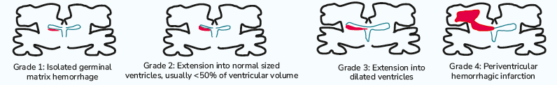
Grade-I IVH: If the hemorrhage occurs only in the subependymal germinal layer (not in the ventricular lumen): Grade-I IVH. Grade-II - IVH: If the hemorrhage extends into the ventricle lumen and occupies 10-50% of the lumen with no ventricular dilatation. Grade-III IVH: If the hemorrhage causes ventricular dilatation and exceeds >50% of lumen. Grade IV-IVH or periventricular hemorrhagic infarction: psilateral parenchyma and venous infarct. In grade IV, venous infarction (IVH) is obstruction of the terminal vein and results in congestion, bleeding, and infarction in the brain parenchyma.
Grades of Ventriculomegaly
- Mild ventriculomegaly: 0.5 to 1 cm dilatation
- Moderate ventilation: 1 to 1.5 cm dilatation
- Severe Ventriculomegaly: > 1.5 cm dilatation
Clinical Presentations
- Asymptomatic or silent bleeding is the most common type of presentation. PCV is found to be abnormal.
- Saltatory bleeding evolves over days or weeks. Lethargy, poor feeding, apnea, toe abnormality, and tight popliteal angle.
- A catastrophic presentation is severe, rare, and life-threatening. Progress over minutes to hours. Apnea, seizures, low SpO2 levels, hypotension, posturing, and can also lead to death.
Also read: Skull Fractures And Traumatic Intracranial Hemorrhage
Investigation and Diagnosis
Transcranial ultrasonography is the IOC for diagnosis. Transcranial ultrasonography is 100% sensitive and 91% specific for detection of IVH with a dimension > 5 mm. All babies born < 32 weeks or weight < 1500 grams should be screened. Babies between 32-34 weeks and associated with RDS/asphyxia/anemia should be screened. First screening is done on D3 or D4 of life; it detects 90% of cases. 50% of detection is done on day 1, 25% of detection is done on day 2, and 15% of detection is done on day 3. If the birth weight is between 500 and 750 grams, D1 screening can be performed. Repeat
- 3 Weeks old (postnatal)
- 36-week postmenstrual age
Progression of IVH
1/3rd of IVH progresses in size: The maximum size increase occurs 3-5 days later.
If on D3 or D4 of screening, IVH is detected, repeated ultrasound is done about 3-5 days later.
.png)
Management of IVH (Intraventricular Hemorrhage)
Supportive care and vigilant monitoring, particularly for the lower grades of IVH.
Supportive Care
- Ensure airway, breathing, and circulation.
- If seizures are present, consider antiepileptic drugs, e.g., phenobarbitone.
- Avoid hypo and hypertension.
- Avoid hypoxia, rapid fluid administration, acidosis, and CO retention. 2
- Avoid and manage pneumothorax.
Monitoring for Post-Hemorrhage Hydrocephalus (PHH)
- Clinical Monitoring
- Alternate-day OFC (occipitofrontal circumference) monitoring.
- If increase in OFC > 2 mm/day, suspicious.
- If increase in OFC > 4mm/every two days, PHH
- If increase > 14mm/week, PHH.
- Watch for signs and symptoms of increased intracranial pressure (ICP) such as bulging anterior fontanelles, separation of cranial sutures (coronal suture first to separate), tone abnormalities, and the sunset sign.
- Alternate-day OFC (occipitofrontal circumference) monitoring.
- Serial Ultrasonography (USG)
Levene's index is assessed, measured from the falx to the lateral wall of the ventricular body. An intervention for PHH might be required if Levene's index exceeds 4mm above the 97th percentile (action line).
- Doppler Ultrasonography (USG)
Performed on the anterior cerebral artery or the middle cerebral artery to
determine the resistance index (RI). RI > 0.85 indicates elevated intracranial
pressure (ICP). RI = 1, Decrease perfusion due to severely elevated ICP, indicate
absent diastolic flow.
Management of PHH
For 1/3rd of PHH patients, spontaneous recovery with conservative treatment. In contrast, 2/3rd of PHH patients require intervention; serial lumbar puncture (LP) is employed in 14% of cases. Shunts are used in 34% of cases. Mortality is recorded in 18% of these cases. According to both the Nelson and AIIMS protocols, there's no established benefit in using agents such as acetazolamide or furosemide for PHH management.
Indications to do Therapeutic Lumbar Punctures
A therapeutic LP should be considered if two or more of the following criteria are met: An increase in OFC (occipitofrontal circumference) of > 14mm per week or an increase of > 2mm per day sustained over 2 weeks. Ventriculomegaly: Levene's index > 97th centile and 4mm above the action line. Signs and symptoms indicating raised intracranial pressure (ICP). A Doppler study reveals a Resistance Index (RI) > 0.85.
Procedure for Tapping CSF: Volume: Extract between 10-20 ml/kg of cerebrospinal fluid (CSF).
Rate: Ensure the tapping rate is below 1 ml/kg/min. If a lumbar puncture is unsuccessful or is deemed unfeasible, a ventricular tap is an alternative, but the risks are: Elevated potential for infections, Loculated hydrocephalus
Indications for VAD or VSG Shunts
VAD stands for Ventricular Access Device, and VSG refers to Ventricular Subgaleal Shunt.
When to Implement VAD or VSG Shunts: Requirement for > 2 lumbar punctures. Or if > 1 ventricular tap is required.
Reasons to Avoid VP Shunts in IVH Preterm Newborns: Higher risk of infection, with an occurrence rate of approximately 15.5%. Higher risk of shunt failure or blockage, in about 17% within three months.
Criteria for Continued Placement of VAD/VSG Shunts: The shunt is not infectious. The rise in OFC (occipitofrontal circumference) stabilizes to normal levels. CSF protein levels < 150 mg/dL. Till a weight of 2.5 kg is attained.
Also read: Retinopathy of Prematurity- Mechanism and Treatment
Download the PrepLadder app now and unlock a 24-hour FREE trial of premium high-yield content. Access Video Lectures, digital notes, QBank, and mock tests for FREE to ace your NEET SS Pediatrics preparation. Elevate your study experience and gear up for success. Start your journey with PrepLadder today!

PrepLadder Medical
Get access to all the essential resources required to ace your medical exam Preparation. Stay updated with the latest news and developments in the medical exam, improve your Medical Exam preparation, and turn your dreams into a reality!
Navigate Quickly
What is IVH?
Site of Origin of IVH
Risk factors of IVH
Nelson 21st Ed-Risk Factors for IVH
Grades of IVH
Grades of Ventriculomegaly
Clinical Presentations
Investigation and Diagnosis
Progression of IVH
Management of IVH (Intraventricular Hemorrhage)
Supportive Care
Monitoring for Post-Hemorrhage Hydrocephalus (PHH)
Management of PHH
Indications to do Therapeutic Lumbar Punctures
Indications for VAD or VSG Shunts
Top searching words
The most popular search terms used by aspirants
- NEET SS Pediatrics Neonatology
- NEET SS Pediatrics Neonatology Preparation
PrepLadder 4.0 for NEET SS
Avail 24-Hr Free Trial