Histology of Skin
Feb 15, 2023

The histology of skin provides insight into the structure and function of the skin, including the different layers and the various cells and tissues involved in maintaining skin health. A good understanding of the histology of skin is important for understanding the anatomic basis of various skin conditions and diseases, such as skin cancer, dermatitis, and acne. Besides, in the NEET PG exam histology of skin is a topic that is frequently tested.
In this blog, we’ve discussed this important anatomy topic briefly for the NEET PG exam preparation. Read on.

Layers of skin:
- The skin's thin outer layer, or epidermis, is the keratinized stratified squamous epithelium. The protective function of skin depends on the epidermis. Dermal papillae are formed by the folding of this epithelium's basal layers. Four different cellular layer types can be found in thin skin, compared to five in thick skin. To learn more about the layers of the epidermis, click here.
- The thicker inner layer known as the dermis. The layer of skin here is made up of connective tissue. Sensation, safety, and thermoregulation all depend on it. Along with sweat glands that open onto the skin's surface and hair in some areas, it also houses nerves, the blood supply, fibroblasts, and other cells. Dermal papillae, which are more noticeable in thick skin, are formed by the folding of the dermis' apical layers.
- A hypodermis. The dermis and this layer below it blend together. Adipose tissue and sweat glands are primarily found there. The generation of triglycerides and vitamin D, as well as other metabolic processes, are carried out by adipose tissue.
Epidermis can have 4 or 5 layers depending on the location it is present in.
Skin has 2 main layers
- Epidermis (upper layer)
- Dermis (lower layer)
Epidermis can have 4 or 5 layers depending on the location it is present in.
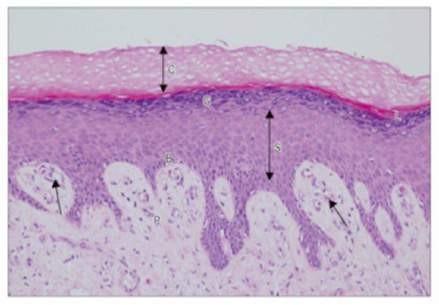
The above skin specimen is taken from the palm (thick skin). Therefore, we can see 5 layers of epidermis.
- Stratum corneum: It has a lot of keratin fibers and dead cells. It is because of these keratin fibers, that this layer has an eosinophilic appearance (pinkish color).
- Stratum lucidum: It is a thin pinkish layer. It is seen only in the areas of thick skin, like the palms and soles. This layer also has dead cells.
- Stratum granulosum: It has kerato-hyaline granules for the formation of keratin filaments.
- Stratum spinosum: It is also known as the ‘Prickle cell layer’ and is the thickest of all layers. It is called so, because the cells in this layer have a spiny or pointed appearance. This spiny appearance is because of the abundance of desmosomes in this layer. The desmosomes in this layer are compromised in diseases like pemphigus vulgaris, which causes skin blisters.
- Stratum basale: It is also known as Stratum germinativum, because this layer has stem cells that keep adding more layers of skin as it sloughs off from the surface. It is a single layer of columnar cells.
The dermis is divided into 2 layers:
- Superficial papillary layer of dermis
- Deep reticular layer of dermis
Superficial papillary layer of dermis -It has some loose areolar connective tissue with some special receptors. In the above skin specimen, we can appreciate a Meissner’s corpuscle.

- Meissner’s corpuscle
It is present in the papillary layer of the dermis. It detects the 2-point discrimination or tactile discrimination.
Also Read: Transverse Section of the Midbrain - NEET PG Anatomy
HISTOLOGY OF THIN SKIN
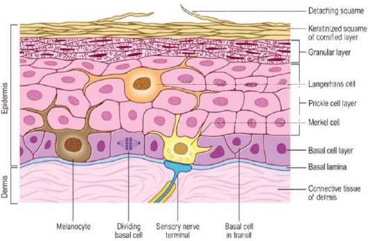
Here, the stratum lucidum is missing. Some special cells seen in stratum basale:
- Melanocytes
- Merkel cells
- Melanocytes:
Derived from neural crest cells (NCCs). It synthesizes melanin, to help our body counter UV radiation from sunlight. It gives multiple processes, which are also reaching the upper layers, like the stratum spinosum.
- Merkel cell:
It is a slowly adapting receptor, for light touch sensations, like holding a pencil.
| Important information: Langerhans cells: These are antigen-presenting cells (APCs). They pick up the antigen and present it to the lymphocytes as they move on towards the lymph nodes. They belong to the monocyte-phagocyte series. They are mostly seen in stratum spinosum. |
Also Read: Development of the Nervous System - NEET PG Anatomy
Fetal Veins, Portal vein formation, Derivatives of the embryonic veins - NEET PG Anatomy
MEISSNER’S CORPUSCLE vs MERKEL CELL
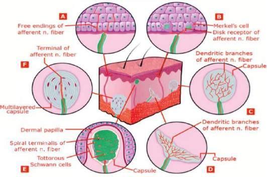
Dermis: It has 2 layers.
- Superficial papillary layer → thin
- Deep reticular layer → thicker; has a larger proportion of connective tissue
| Meissner’s corpuscle (E) | Merkel cell (B) |
| It is an encapsulated receptor, with multiple stacks of cells, for 2-point discrimination. | It is a slowly adapting receptor, it detects light touch sensations. |
| It is a rapidly adapting receptor | It is a slowly adapting receptor |
| It is seen at the dermo-epidermal junction, in the papillary layer of dermis. | It is seen in stratum basale. |
Important Information:
- Braille is read with the help of Meissner’s corpuscle. This is because it is a rapidly adapting receptor, which makes reading braille easier.
- However, the most precise receptor to read Braille is → Merkel cell > Meissner’s corpuscle.
- But since the Merkel cell is a slowly adapting receptor, it helps in identifying the character but does not help us in reading the sentences.
- Meissner’s corpuscle can also help in detecting low frequency vibrations.
- Pacinian corpuscle (F):
- It is present in the deep dermis.
- It helps in detecting pressure and high frequency vibrations.
- It is a rapidly adapting receptor.
- Ruffini receptor (D):
- It is present in the deep dermis.
- It helps in detecting the dermal stretch, when there is a stretch on the skin.
- It is a slowly adapting receptor.
Also Read:
Gametogenesis - Definition, Stages and Types - NEET PG Anatomy
Inguinal Canal & Spermatic Cord - NEET PG Anatomy
Development of Skull - NEET PG Anatomy
To study histology of skin in detail, download the PrepLadder app and find detailed video lectures and study notes covering the topic.

PrepLadder Medical
Get access to all the essential resources required to ace your medical exam Preparation. Stay updated with the latest news and developments in the medical exam, improve your Medical Exam preparation, and turn your dreams into a reality!
Navigate Quickly
Layers of skin:
Skin has 2 main layers
HISTOLOGY OF THIN SKIN
MEISSNER’S CORPUSCLE vs MERKEL CELL
Top searching words
The most popular search terms used by aspirants
- Anatomy Important Topics
- NEET PG Anatomy