Neural Crest Cells and Germ Layer Derivatives: The Complete NEET PG Guide
Dec 4, 2025

The Question That Stumps Most Candidates
A newborn shows hypocalcemic convulsions, coarctation of the aorta, cleft palate, and no thymic shadow on the chest X-ray. DiGeorge syndrome is suspected by the pediatrician.
The examiner must understand which embryological structure produces this particular constellation when it is defective.
This is precisely the gap we need to fill if your response was "neural crest cells", but you're not entirely aware why the thymus, parathyroids, and cardiac outflow tract are all harmed simultaneously.
Questions on embryology stress comprehension above memorisation. When you know which structure gives birth to what, you may foresee which defects will cluster together. That's the difference between memorising a list and genuinely reasoning through a narrative.
NEET PG EXAM PATTERN
Germ layer derivatives appear in 2 to 4 questions annually. Sometimes, as a direct question (X is derived from which germ layer?), it is embedded in clinical vignettes about congenital anomalies. Neural crest derivatives are particularly high-yielding.
The Short Answer
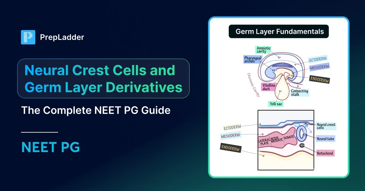
Germ layers, i.e ectoderm, mesoderm, and endoderm, are the three primary tissue layers established during gastrulation in week 3. Every structure in your body traces back to one of these three.
The dorsal aspect of the neural tube gives rise to a unique migratory cell population known as neural crest cells. They travel widely and produce structures all across the body, including melanocytes, peripheral ganglia, adrenal medulla, craniofacial bones, and portions of the heart outflow system, despite coming from ectoderm.
The main insight: Neural crest cells behave nearly like a fourth germ layer since they create connective tissues like bones and cartilage that you'd intuitively expect from mesoderm.
Also Read: Nymphomania: Causes, Symptoms, Risk Factors, Diagnosis, Treatment
Gastrulation: Where It All Begins
By the third week, gastrulation leads the bilaminar disc to transform into a trilaminar disc. Through the primitive streak, epiblast cells migrate inward:
Some are still superficial, such as the ectoderm
Some move to the mesoderm, which is in the centre.
Some move the endoderm and hypoblast.
The epiblast is the source of all three germ layers. The hypoblast contributes only to extraembryonic structures, not to the embryo itself.
Access NEET PG Previous Year Question Papers PDF – Free Download
Ectoderm: The Outer Layer
Ectoderm has two major divisions based on what happens during neurulation:
Surface ectoderm, which stays on the outside, and Neuroectoderm, which folds inward to form the neural tube.
Surface Ectoderm Derivatives
Structures that interact with the outside environment are formed by surface ectoderm:
Skin and appendages:
- Epidermis (note: dermis is NOT from ectoderm)
- Hair follicles
- Nails
- Sweat glands
- Sebaceous glands
- Mammary glands
Sensory structures from placodes:
- Lens of the eye (derived from lens placode)
- Inner ear membranous labyrinth (from otic placode)
- Olfactory epithelium (from nasal placode)
Oral ectoderm derivatives:
- Enamel of teeth - the only ectodermal component of a tooth.
- Anterior pituitary - from Rathke's pouch, an ectodermal diverticulum from the roof of the stomodeum
- Parotid gland
The anterior pituitary question is a common one. Rathke's pouch develops upward from the mouth ectoderm. From the floor of the diencephalon (neuroectoderm), the posterior pituitary descends. Two embryological origins, one gland. The suprasellar position of craniopharyngiomas is explained by their origin from remnants of Rathke's pouch.
Neuroectoderm Derivatives
When the neural plate folds to form the neural tube, it gives rise to the entire central nervous system:
- Brain (cerebrum, cerebellum, brainstem)
- Spinal cord
- Retina and optic nerve
- Posterior pituitary (neurohypophysis)
- Pineal gland
- CNS glial cells: oligodendrocytes and astrocytes
What is not derived from neuroectoderm: Peripheral nerves, Schwann cells, dorsal root ganglia, and autonomic ganglia. All of them are neural crest derivatives, which examiners frequently test.

Neural Crest: The Migratory Population
This is the stage at which embryology truly shines.
Cells at the contact between the neural tube and surface ectoderm detach and migrate broadly throughout the embryo as the neural tube closes. These neural crest cells inhabit regions from head to pelvis, growing into a surprising range of tissues.
Despite its ectodermal origin, the neural crest is frequently referred to as "ectomesenchyme" since these cells produce bone, cartilage, and connective tissue—structures typically associated with mesoderm. This dual nature is precisely why they're checked so routinely.
Also Read: Modes of Ventilation: Types and Uses
Additional Neural Crest Derivatives
- Pharyngeal arch cartilages.
- Ciliary body and stroma of the iris.
- Corneal stroma and endothelium.
- Aorticopulmonary septum.
- Parafollicular cells (C-cells) of the thyroid
- Connective tissue of the thymus and parathyroids
The Face Is Neural Crest Territory
The craniofacial skeleton is predominantly neural crest-derived. This includes:
- Maxilla and mandible
- Zygomatic bones
- Nasal bones
- Frontal bone
The occipital bone, however, is primarily paraxial mesoderm (somite-derived).
When Neural Crest Migration Fails: DiGeorge Syndrome
Now the opening case makes sense.
Neural crest cells migrate into pharyngeal arches 3 and 4 during development. These neural crest cells contribute to:
- Thymus
- Parathyroid glands
- Aortic arch derivatives and cardiac outflow tract
- Craniofacial structures
In DiGeorge syndrome (22q11.2 deletion), neural crest migration into these arches is defective:
- Thymic hypoplasia → T-cell immunodeficiency
- Parathyroid hypoplasia → hypocalcaemia → seizures
- Defects in cardiac outflow: Fallot's tetralogy, truncus arteriosus, and interrupted aortic arch
- Deformities of the face: cleft palate, low-set ears
One embryological lesion. One clinical syndrome. This is pattern recognition, not memorisation.
Other Neural Crest-Related Disorders
Condition Neural Crest Derivative Affected Hirschsprung disease Enteric ganglia are absent in the distal colon Waardenburg syndrome Melanocytes (white forelock, heterochromia) Neuroblastoma Sympathetic ganglia or adrenal medulla Treacher Collins syndrome First and second pharyngeal arch neural crest Piebaldism Melanocytes
Also Read: Pre-Anesthetic Checkup (PAC)
Mesoderm: The Middle Layer
Mesoderm organises into three different sections, each with particular derivatives.
Paraxial Mesoderm (Somites)
The neural tube is flanked by segmented pieces of paraxial mesoderm called somites. Each somite separates into three components:
Sclerotome (ventromedial):
- Vertebral column
- Ribs
- Occipital bone (posterior skull base)
Myotome (central):
- Skeletal muscles of the trunk and limbs
- Muscles retain segmental innervation because each myotome keeps its original spinal nerve
Dermatome (dorsolateral):
- Dermis of the skin over the back
Note: Dermis of the face and anterior neck is neural crest, not mesoderm. Lateral plate mesoderm makes up the dermis of the limbs and anterior trunk.
Intermediate Mesoderm
This strip between paraxial and lateral plate mesoderm generates the urogenital system:
- Kidneys (pronephros -> mesonephros -> metanephros)
- Ureters
- Gonads (ovaries, testes)
- Adrenal cortex
The cortex is an intermediate mesoderm, and the adrenal gland has two origins. The medulla is neural crest. This is why pheochromocytoma (neural crest tumour) and adrenocortical carcinoma are biologically separate neoplasms.
Lateral Plate Mesoderm
The lateral plate separates into two layers with the intraembryonic coelom between them:
Somatic (parietal) layer:
- Bones of limbs (appendicular skeleton)
- Dermis of limbs and body wall (except back)
- Parietal serous membranes (parietal pleura, parietal peritoneum, parietal pericardium)
Splanchnic (visceral) layer:
- Heart and all blood vessels
- Smooth muscle and connective tissue of the gut wall
- Visceral serous membranes
- Spleen
Mesoderm Summary Table
Region Key Derivatives Paraxial (Somites) Vertebrae, ribs, skeletal muscles, and dermis of the back Intermediate Kidneys, ureters, gonads, adrenal cortex Lateral Plate: Somatic Limb bones, parietal serous membranes Lateral Plate: Splanchnic Heart, blood vessels, gut smooth muscle, spleen
Endoderm: The Inner Layer
The endoderm is your innermost germ layer, and it's responsible for producing the epithelial lining of the gut tube and anything that buds off from it. The word "epithelial lining" is crucial to keep in mind because endoderm only produces the inner lining; mesoderm and neural crest, respectively, produce the gut's muscular wall, blood supply, and nerve plexuses.
So when you think about the GI tract, from the oropharynx all the way down to the upper anal canal, that entire epithelial lining is endodermal in origin. The respiratory system follows the same rationale since it develops as an extension from the foregut called the laryngotracheal diverticulum.
This means the epithelium lining your larynx, trachea, bronchi, and alveoli is all endoderm-derived. But here's where students typically trip up – the cartilages that support the larynx, like your thyroid, cricoid, and arytenoid cartilages, are not endodermal.
Those come from neural crest cells as pharyngeal arch descendants. Therefore, the lining is endoderm, while the surrounding structural support has an entirely distinct embryological genesis.
Glandular Derivatives
When it comes to glandular derivatives, endoderm produces the functional parenchyma of several important organs that bud out from the foregut. This comprises the exocrine and endocrine parts of the pancreas, thyroid follicular cells, liver hepatocytes, and gallbladder epithelium. However, there's always a catch with embryology – the C-cells of the thyroid, which make calcitonin, are actually neural crest in origin, not endodermal. This distinction comes up regularly in tests, so keep it in mind.
The pharyngeal pouches are endodermal outpouchings that arise between the pharyngeal arches, and each one gives rise to unique structures. The tympanic cavity and Eustachian tube epithelium develop from the initial pouch. The palatine tonsil's epithelium in the tonsillar fossa is formed by the second pouch. The inferior parathyroid glands and the thymus epithelium are located in the third pouch. The fourth pouch gives birth to the superior parathyroid glands and the ultimobranchial body.
Now here's a famous conundrum that confuses everyone - why is the inferior parathyroid generated from the third pouch but placed below the superior parathyroid from the fourth pouch? The answer lies in migration. The third pouch derivatives, notably the thymus and inferior parathyroid, move caudally during development.
The inferior parathyroid is effectively dragged by the thymus as it descends all the way into the mediastinum. Meanwhile, the fourth pouch derivatives stay basically put. So even though the third pouch is anatomically higher up initially, its derivatives end up lower in the final arrangement.
One more topic that trips folks up is the origin of C-cells. The ultimobranchial body itself comes from the fourth pharyngeal pouch, making it endodermal. But the parafollicular C-cells within it actually arise from neural crest cells that migrate into this structure. So the pouch is endoderm, but the cells that settle there and create calcitonin are neural crest – a small but significant distinction.
The Master Comparison Table
Structure Germ Layer Origin Epidermis Surface ectoderm Dermis of face/anterior neck Neural crest Dermis of the back Paraxial mesoderm (dermatome) Dermis of limbs/trunk Lateral plate mesoderm CNS neurons Neuroectoderm CNS glia (astrocytes, oligodendrocytes) Neuroectoderm PNS neurons (all ganglia) Neural crest Schwann cells Neural crest Skeletal muscle Mesoderm (myotome) Smooth muscle Mesoderm (splanchnic) Cardiac muscle Mesoderm (splanchnic) Facial skeleton Neural crest Occipital bone Paraxial mesoderm Vertebrae and ribs Paraxial mesoderm (sclerotome) Limb bones Lateral plate mesoderm Adrenal cortex Intermediate mesoderm Adrenal medulla Neural crest Anterior pituitary Surface ectoderm (Rathke's pouch) Posterior pituitary Neuroectoderm Thyroid follicular cells Endoderm Thyroid C-cells Neural crest Parathyroid epithelium Endoderm Enamel Surface ectoderm Dentin Neural crest (odontoblasts) Pulp of the tooth Neural crest Kidney Intermediate mesoderm GI tract epithelium Endoderm GI tract smooth muscle Splanchnic mesoderm Enteric ganglia Neural crest Liver hepatocytes Endoderm Blood vessels Mesoderm Melanocytes Neural crest Lens of the eye Surface ectoderm Retina Neuroectoderm Spleen Mesoderm (splanchnic) Pia and arachnoid mater Neural crest Dura mater Mesoderm
High-Yield Facts for NEET PG
- The neural crest, which forms bones, cartilage, and connective tissue in the head and neck, develops from ectoderm but acts like mesoderm.
- Adrenal gland = dual origin. Cortex from intermediate mesoderm. Neural crest medulla.
- Rathke's pouch (surface ectoderm) is the anterior pituitary. Posterior pituitary = infundibulum (neuroectoderm). Rathke's pouch remnants give rise to craniopharyngioma.
- The only ectodermal derivative found in teeth is enamel. Dentin, cementum, and pulp are neural crest.
- All three muscle types are mesodermal — skeletal (paraxial), smooth (splanchnic), cardiac (splanchnic).
- Laryngeal cartilages are not mesoderm but rather neural crest, which is derived from the pharyngeal arch. Laryngeal epithelium is endoderm.
Frequently Asked Questions
What distinguishes neuroectoderm from neural crest?
Neuroectoderm creates the neural tube and becomes the central nervous system – brain, spinal cord, retina. PNS ganglia, melanocytes, craniofacial bones, and the adrenal medulla are examples of peripheral structures formed by neural crest cells that migrate from the dorsal neural tube. Neural crest migrates peripherally, but neuroectoderm remains central.
Why do so many different organs get harmed by DiGeorge syndrome?
The thymus, parathyroids, heart outflow tract, and facial structures all require neural crest cell contribution throughout development. In DiGeorge syndrome (22q11.2 deletion), neural crest migration into pharyngeal arches 3 and 4 is aberrant.
Multiple organ issues emerge from a single embryological defect; these anomalies are related by developmental origin rather than function.
Why is the brain generated from mesoderm, and the adrenal medulla from neural crest?
The adrenal cortex is derived from intermediate mesoderm, just as the kidneys and gonads. During development, neural crest cells travel into the developing cortex and settle in the core, forming the medulla. These neural crest cells differentiate into chromaffin cells, which are essentially modified postganglionic sympathetic neurones.
This dual genesis explains why cortical and medullary tumours are physiologically unique.
How do I remember which structures are neural crest?
It's probably neural crest if you ask yourself, "Did this structure require cells to migrate a long distance during development?" Melanocytes travel from the neural tube to the skin. The entire length of the intestine is home to enteric ganglia.
Neural crest cells migrate into pharyngeal arches to generate craniofacial bones. The migration principle helps more than rote memorisation.
Are the cartilages of the pharyngeal arches derived from the neural crest or mesoderm?
Neural crest cells migrate into the pharyngeal arches to generate the cartilages, which include cartilages of the larynx (thyroid, cricoid, and arytenoid). Students frequently confuse "cartilage" with "mesoderm," which is a common misconception. However, it's crucial to remember that the neural crest is where craniofacial and pharyngeal arch cartilages originate.
Download the PrepLadder app now and unlock a 24-hour FREE trial of premium high-yield content. Access Video Lecturesalso in हिंglish, digital notes, QBank, and Mock Tests for FREE to ace your NEET PG preparation. Elevate your study experience and gear up for success. Start your journey with PrepLadder today!

PrepLadder
Access all the necessary resources you need to succeed in your competitive exam preparation. Stay informed with the latest news and updates on the upcoming exam, enhance your exam preparation, and transform your dreams into a reality!
Navigate Quickly
The Question That Stumps Most Candidates
Gastrulation: Where It All Begins
Access NEET PG Previous Year Question Papers PDF – Free Download
Ectoderm: The Outer Layer
Surface Ectoderm Derivatives
Neuroectoderm Derivatives
Neural Crest: The Migratory Population
Additional Neural Crest Derivatives
The Face Is Neural Crest Territory
When Neural Crest Migration Fails: DiGeorge Syndrome
Other Neural Crest-Related Disorders
Mesoderm: The Middle Layer
Paraxial Mesoderm (Somites)
Intermediate Mesoderm
Lateral Plate Mesoderm
Mesoderm Summary Table
Endoderm: The Inner Layer
Glandular Derivatives
The Master Comparison Table
High-Yield Facts for NEET PG
Frequently Asked Questions
Top searching words
The most popular search terms used by aspirants
- Anatomy Important Topics
- Medical PG Anatomy
- NEET PG Anatomy Preparation