Genitourinary Radiology- Micturating/Voiding Cystourethrography
Apr 26, 2023

Genitourinary radiology involves the imaging techniques used to diagnose and manage a variety of genitourinary disorders. The genitourinary system includes the organs of the urinary tract and the reproductive system, which are susceptible to a wide range of conditions, including infections, tumors, and congenital abnormalities.
Additionally, the topic of genitourinary radiology is relevant to many different medical specialties, including urology, gynecology, oncology, and radiology. Therefore, developing a good understanding of this topic is crucial.
In this blog we’ll cover horseshoe kidney , micturating/voiding cystourethrography, breast imaging. Read on.

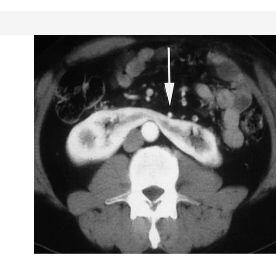
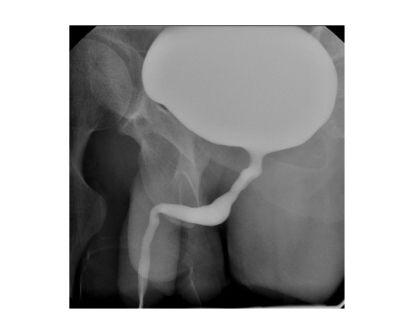
Ureterocele
- Cystic dilatation in distal ends of ureter: “cobra / Adder head appearance”
- Types
- Simple
- Ectopic
- Seen with duplicated collecting system (upper pole collecting system)
Horseshoe Kidney

- Most common fusion Anomaly in kidney
- Inferior poles of kidney are medially located (B/L) and are fused : “flower vase/shaking hand calyces”
- Fused segment is referred as “Isthmus”
- CECT
- Isthmus seen
- Inferior mesenteric artery is in relation to isthmus (Blocking the ascend of kidney) : L3 level
- Complications
- Ureteric obstruction [due to medially oriented ureters]
- UTI / Stasis / calculi
- Higher incidence of trauma
- Ureteric obstruction [due to medially oriented ureters]
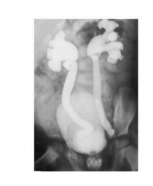
Duplex Collecting System
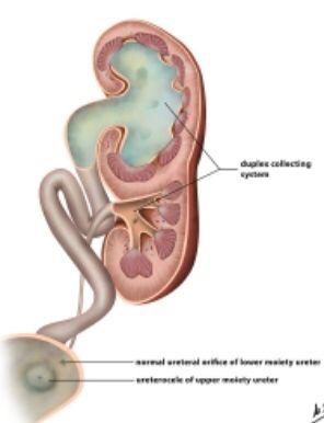
- “Drooping Lilly sign” in case of Duplicating collecting system
- Weight – Meyer law
- Upper pole and lower pole collecting system has their own ureter
- Upper pole: Opens inferomedially in relation to trigone
- Upper pole collecting system more prone for
- Obstruction
- Ectopic insertion
- Ureterocele formation
- Lower pole opens superolaterally; has complication of vesicoureteral reflux
Polycystic kidney disease (Autosomal dominant)
- “Spider leg sign”: Splaying of pelvicalyceal system Belatedly
- “Swiss cheese appearance”: In nephrographic phase when the renal cortex has contrast it shows filling defect.
- USG: Entire kidney has multiple large cyst Bilaterally

Medullary sponge kidney
Paint brush appearance
- “Paint brush appearance”/ “Bouquet of flowers”: Calcification in medullary pyramids along with the contrast that is getting excreted
Retroperitoneal fibrosis
- Both ureters are medially deviated referred as “Maiden waist sign”
- Causes
- Primary / Ormond disease
- 2° (secondary)
- IgG 4 Related disease
- Post radiotherapy
- Drug like methysergide
Retrocaval Ureter

- Kidney is hydronephrotic: proximal ureter is dilated; acute curvature giving ; “ J shaped / hook- like ureter”
- Ureter passer Behind IVC (anomalous)
- It is because of abnormal IVC Embryology that ureter goes behind IVC.
Papillary necrosis

- Necrotic sluff of papillae within renal calyces referred as “signet ring sign ” / “Ball on tee sign” / “Lobster claws sign”
- Causes
- Associated with NSAIDS
- Renal TB
- Sickle cell Anemia
- DM
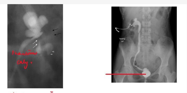
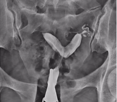
Retrograde urethrogram
- Retrograde urethrogram
- IOC for anterior urethra
- IOC for urethral Injury
Important Information
- Anterior segment of male urethra formed by
- Penile urethra
- Bulbar urethra
- Posterior segment of male urethra formed by
- Prostatic
- Membranous
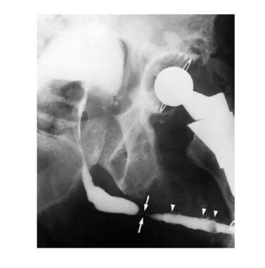
- Peno-bulbar stricture
- Penile urethra has irregularities; at the junction of penile & Bulbar urethra there is complete narrowing (stricture)
- Retrograde urethrogram : IOC (for anterior urethral strictures)
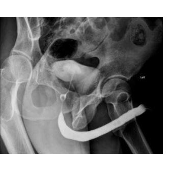
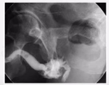
Micturating/Voiding cystourethrography
- Micturating / voiding cystourethrography
- Bladder is catheterized with foley then 300- 400 ml of diluted contrast is injected (stop injecting when bladder in full)
- Under x-ray patient is asked to void
- IOC:
- Vesicoureteral reflux (VUR)
- Posterior urethral value (PUV) (anterior ureter not getting contrast)
- Grade 5 vesico ureteric reflux
- Along with bladder gross distention of ureter, gross hydronephrosis, ballooning of calyces
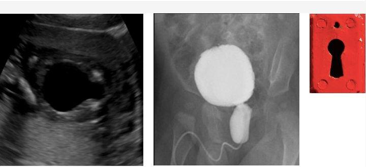
- Posterior urethral value
- “Key Hole Sign”: Dilated bladder and posterior urethra
- IOC: MCU
Bladder Appearances in Different Conditions
Neurogenic Bladder Tear drop Fetal skull appearance
- Vertically oriented “Christmas tree” bladder with multiple diverticulation; VUR (+) →Neurogenic Bladder
- Bladder is compressed; “Teardrop/pear shaped” appearance → extrinsic compression [due to masses like pelvic lipomatosis, pelvic hematoma]
- “Fetal skull appearance”; Bladder wall showing calcification → schistosomia infection
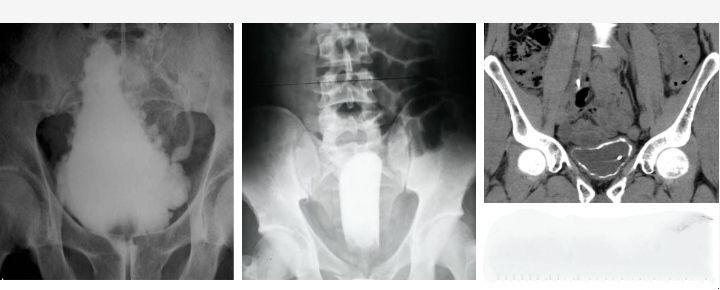
Genitourinary TB
Thimble bladder
- MC affected : Kidney
- Hematogenous route
- Earliest finding in IVP: Moth eaten calyces (irregular calyces)
- Most sensitive investigation: IVP
- DOC: CT urography
- End stage we can see: Various Strictures, very irregular cycles
- Phantom calyx: When calyx is not seen
- Entire ureter narrows referred as “pipe stem ureter”
- Bladder capacity ↓: Thimble Bladder
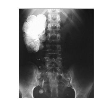
- End stage
- Kidney is calcified: Putty kidney (with absolute loss of function)
- Therefore, known as Auto nephrectomy
Radiology Related Articles:
| Why is Radiology the most preferred branch? | RADIOTHERAPY Types| Cancer treatment with Radiation: NEET PG Radiology | Musculoskeletal Radiology: Bone Tumors |
Also watch a related video on Genitourinary By Dr. Nikita Nanwani:
Emphysematous pyelonephritis
- History of patient in sepsis
- All trapped in renal fossa
- Polymicrobial infection mainly of gram-Negative organisms.
- Poor prognosis
- To do pig tail drain; if sepsis not controlled then do nephrectomy
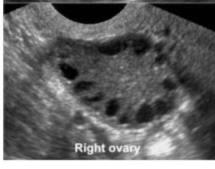
PCOS
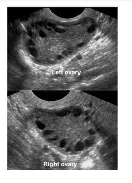
- Polycystic ovarian Syndrome
- Ovaries increased in volume :>10 CC [Bulky ovaries]
- Multiple peripheral follicles: 2-8mm; which are more than 12 in number
- No dominant following (No ovulation)
- Central Stroma has raised volume and echogenicity: Most specific feature of PCOS
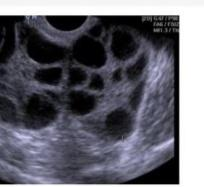
OHSS vs Theca Lutein Cyst
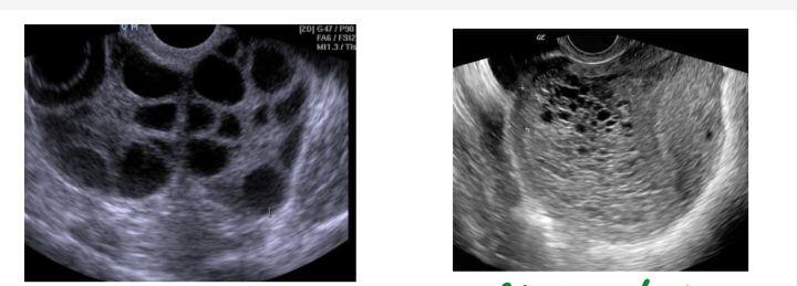
- Multiple large follicles in Both ovaries
- Two possibilities,
- OHSS [if any History of IVF, patient Comes in hypotensive state, pleural effusion, Ascites]
- Theca Lutein cyst [seen in relation with molar pregnancy]
- Two possibilities,
Important Information
- Molar Pregnancy
- Cluster of grapes appearance
- Snowstorm appearance
Anomalies
- Anencephaly
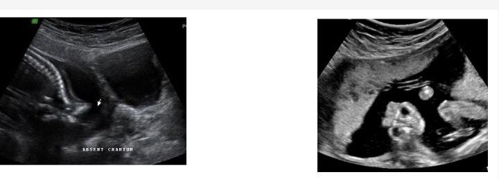
- Frog – eye sign
- Earliest anomaly that can be detected [11th week scan ]
- Acrania
- “Frog - eye sign" /“ Mickey Mouse sign": Eye is very prominent due to absent of Skull ;dysplastic Brain
Gastroschisis Omphalocele
|
Gastroschisis |
Omphalocele |
|
|
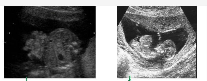
Twin pregnancy
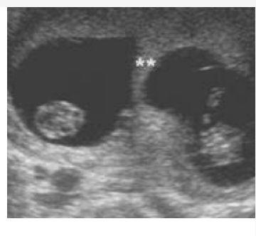
DCDA
MCDA
- Twin peak sign / Lambda sign: Dichorionic Diamniotic (intertwin membrane > 2mm)
- T sign: Monochorionic diamniotic (intertwin membrane < 2mm)
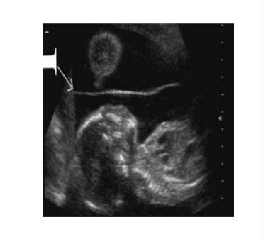
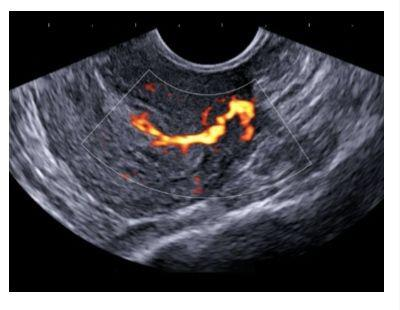
Endometrial polyp

- Feeding vessel sign on doppler: Hallmark of Endometrial polyp
Breast Imaging

Cc- Craniocaudal view MLO- Mediolateral oblique vies
- More breast tissue Covered in : MLO
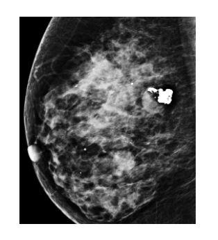
- Fibroadenoma
- Coarse calcification referred as "Popcorn Sign"
- Particularly in Involuting Phase
- Important information
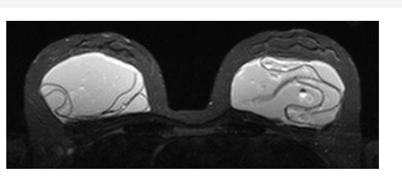
- IOC for breast Implant: MRI
- Intra capsular rupture of Breast Implant
- Linguine Sign/Tear drop Sign / keyhole sign
Previous Year Question
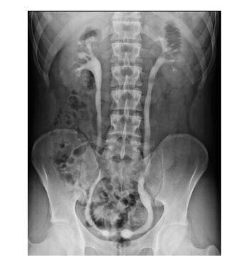
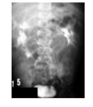
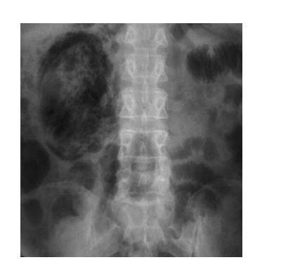
Q. 35-year-old patient comes to the emergency room with vomiting and colicky abdominal pain. On investigation the following image is obtained. Likely diagnosis is? (FMGE Dec 2020)

A. Pancake Kidney
B. Ectopic Kidney
C. Horseshoe Kidney
D. Crossed fused ectopia
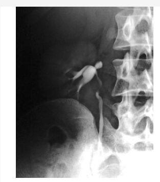
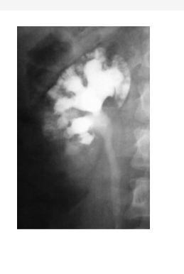
Q. Identify the investigation and Diagnosis? (NEET JAN 2020)

A. MCU, Bulbar Urethral Stricture
B. RGU, Prostatic Urethral Stricture
C. RGU, Bulbar Urethral Stricture
D. MCU, Prostatic Urethra Stricture
Q. Diagnosis is based on the image shown? (FMGE Dec 2020)
A. PCOD
B. OHSS
C. Ovarian Cyst
D. Theca Lutein Cyst
Q. A 35 year old female presents to you with a history of nausea and vomiting. She is undergoing IVF treatment, What is the likely diagnosis? (NEET Jan 2020)
A. PCOD
B. Theca-lutein Cyst
C. OHSS
D. Mucinous cystadenoma
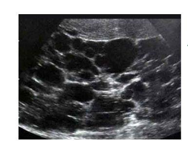
Q A 45-year-old female present with abnormal uterine bleeding. USG is shown here. What is the likely diagnosis ? (INI CET 2020)
A. Ovarian dermoid
B. Endometrial hyperplasia
C. Endometrial carcinoma
D. Endometrial polyp
Q The Most appropriate View for Mammography? (FMGE 2021)
A. Bird’s eye View
B. Spot Compression View
C. Medio lateral oblique
D. Lateral
Q A 30 year old with motor vehicle accident presented to the causality with pelvic fracture. His vitals are stable but he is unable to pass urine. He has blood at the urethral meatus. An RGU was performed as shown below. What is the most likely site of urethral injury? (NEET PG - 2022)
A. Penile urethral
B. Membranous urethra
C. Spongy urethra
D. Bulbar urethra
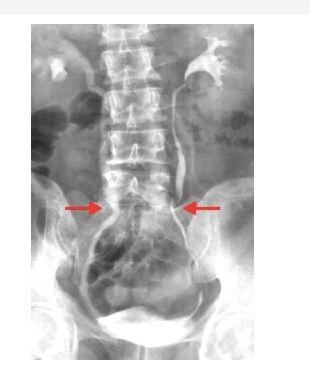
Q. A delayed intravenous urogram of the patient is shown. What is the most likely diagnosis? (NEET PG - 2022)

A. Staghorn calculus
B. Putty Kidney
C. Pelviuretic junction obstruction
D. Renal Cyst
Q. A 22 year old female with primary infertility underwent HSG examination and shown below. What is the diagnosis ? (NEET PG - 2022)
A. Unicornuate uterus
B. Bicornuate uterus
C. Septate uterus
D. Uterus diadelphys
To study this topic in detail along with other high-yield radiology topics, download the PrepLadder app and discover engaging video lectures by expert faculty.

PrepLadder Medical
Get access to all the essential resources required to ace your medical exam Preparation. Stay updated with the latest news and developments in the medical exam, improve your Medical Exam preparation, and turn your dreams into a reality!
Navigate Quickly
Ureterocele
Horseshoe Kidney
Duplex Collecting System
Polycystic kidney disease (Autosomal dominant)
Medullary sponge kidney
Retroperitoneal fibrosis
Retrocaval Ureter
Papillary necrosis
Retrograde urethrogram
Micturating/Voiding cystourethrography
Bladder Appearances in Different Conditions
Neurogenic Bladder Tear drop Fetal skull appearance
Genitourinary TB
Thimble bladder
Emphysematous pyelonephritis
PCOS
OHSS vs Theca Lutein Cyst
Anomalies
Twin pregnancy
Endometrial polyp
Breast Imaging
Previous Year Question
Top searching words
The most popular search terms used by aspirants
- NEET PG Radiology
- NEET PG Strategy