Lasers in eye: Types, Needs, And Applications : NEET PG Ophthalmology
Apr 11, 2023

To truly master Ophthalmology, you must know in depth everything about lasers used on the eyes including their needs, types and applications. This is an important topic for Ophthalmology paper.
Read this blog post carefully to know everything about lasers in the eye for your Ophthalmology paper and ace your NEET PG preparation.

Types of Lasers
Lasers for eyes, based on their mechanisms, are classified into three types:
- Photo disruptive laser
- Photocoagulative laser
- Photo ablative laser
Photodisruptive Lasers
- Photo disruptive lasers are used for cutting a hole in the eye, such as Nd: YAG.
Nd: YAG
- Nd: YAG is used for cutting the posterior capsule, and this procedure is called posterior capsulotomy. So, one application of this laser is laser posterior capsulotomy.
- It cannot be applied to the retina as there will be a retinal hole. Another indication is its use in the peripheral eye, known as peripheral iridotomy. So, the two major indications for Nd: YAG are posterior capsulotomy and peripheral iridotomy.
Femto-Laser
- Femto-laser is another photo disruptive laser. Femto-laser is used in cataract surgery for capsular axis, lens fragmentation, etc., and refractive surgery. The basis for all refractive surgery, especially laser-assisted ones, is to alter the cornea's curvature, which is called keratomileusis. It is known that the more curvature, more is the refractive power which is myopia. If a patient already has myopia, more curvature is unwanted. The correct way would be to flatten the central cornea. In the case of hypermetropia, the correct way would be to bulge the central cornea. The altering of curvature of the cornea as per the need of the patient is called keratomileusis. The common surgery is LASIK surgery. In LASIK surgery, Femto-laser is used to raise the flap. The second application of Femto is the SMILE procedure, where we focus the Femto laser directly on the stroma, cutting a piece of stroma as per the requirement, making a small incision, and removing that piece. SMILE stands for Small Incision Lenticule Extraction.
Photocoagulative Lasers
- This is the second type of laser. One of its applications is on the trabecular meshwork or trabeculoplasty in open-angle glaucoma.
- The second application is on the vascular diseases of the retina.
- The names of Photocoagulative lasers are:
- Argon
- Diode
- Double frequency Nd: YAG (the wavelength here is half of the Nd: YAG).
- Simple Nd: YAG is photo disruptive, but in case of double frequency, it becomes Photocoagulative.)
Photo ablative Lasers
- Photo ablative lasers primarily refer to Excimer lasers. They are of many types
- Xenon Chloride
- Xenon Fluoride
- Argon Fluoride
- Argon Fluoride is used for the eye, and the indication for this is refractive surgery.
What is the wavelength of the laser used in LASIK surgery?
The wavelength of Argon Fluoride is 193 nm.
- The wavelengths of the rest of the lasers are as follows:
- The wavelength of Nd: YAG is an infrared range of 1064 nm.
- The wavelength of the Femto-laser is 1054 or 1053 nm.
- The wavelength of Photocoagulative lasers are as follows:
- Argon: 514 nm
- Diode: 780 nm to 850 nm
- Double frequency Nd: YAG: 832 nm
Ophthalmology Related articles:
Laser Application
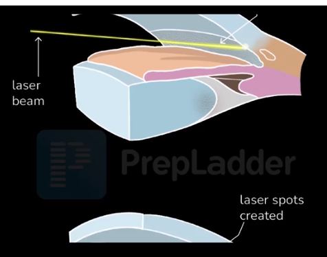
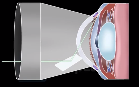
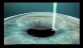
- The laser is applied to the trabecular meshwork, and the laser spots are visible. The laser is focused through the lens.
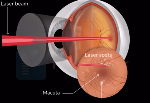
- In the above slide, a laser beam is focussed on the retina in case of Photocoagulative procedures, such as diabetic retinopathy, CRVO, sickle cell, etc.
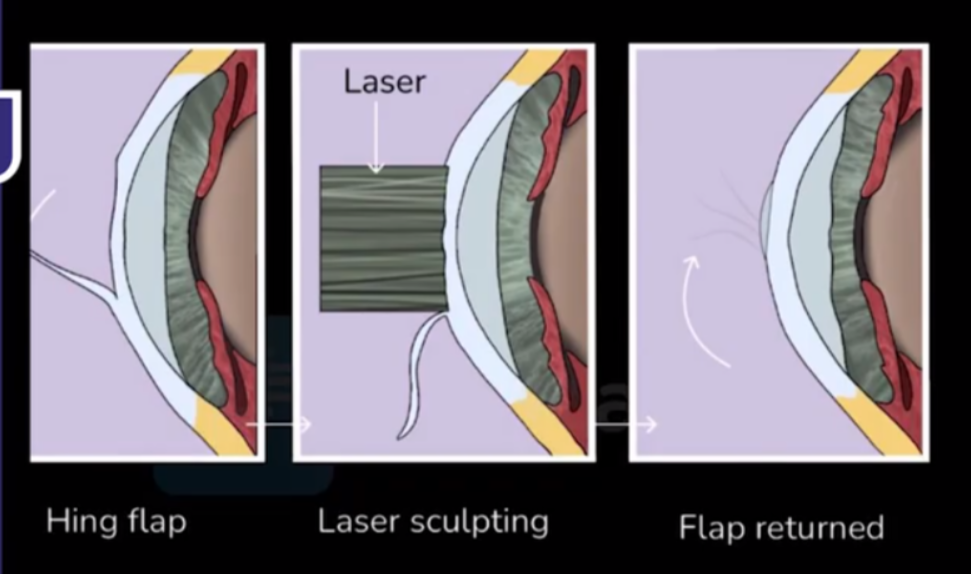
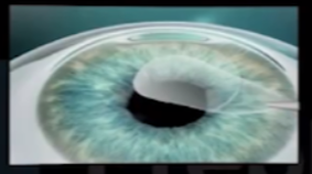
- In this image, it can be seen how LASIK surgery works. There is a flap, and the laser is applied to the rest of the retina. There is sculpting, and the flap returns to its position.
The treatment steps
1. Step 1: Lenticule creation
- A thin tenticule and small incision are created inside the intact cornea.
2. Step 2: Lenticule removal
- The lenticule is removed through the incision with minimal disruption to the corneal biomechanics.
3. Step 3: Impairment is corrected
- Removing the lenticule changes the shape of the cornea, thereby achieving the desired refractive correction.
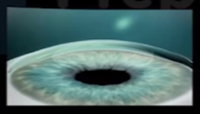
- In the above diagram, the SMILE procedure is being done. In step 1, there’s a creation of the lenticule by the laser; secondly, the lenticule is removed. In the third step, there’s a correction of impairment by changing the shape of the cornea. Lenticule is a piece of the corneal stroma cut by the Femto laser and then removed.
Also Read:
And that is it! You have now covered everything you need to know about Lasers in the Eye for your Ophthalmology paper preparation. For more interesting and informative posts like these, download the PrepLadder App keep following our blog.

PrepLadder Medical
Get access to all the essential resources required to ace your medical exam Preparation. Stay updated with the latest news and developments in the medical exam, improve your Medical Exam preparation, and turn your dreams into a reality!
Navigate Quickly
Types of Lasers
Photodisruptive Lasers
Nd: YAG
Femto-Laser
Photocoagulative Lasers
Photo ablative Lasers
What is the wavelength of the laser used in LASIK surgery?
Laser Application
The treatment steps
Top searching words
The most popular search terms used by aspirants
- NEET PG Ophthamology
- NEET PG Preparation