Layers of Eye : NEET PG Ophthalmology
May 17, 2023

The three layers, namely the sclera, the choroid (the posteriorly extending choroid; extending anteriorly as ciliary body and iris), and the innermost layer which is the retina, can also be seen. Volume of eyeball /globe: 6 ml. Volume of each orbit: 30 ml It is Encased in Tenon’s capsule, suspended by orbit and supported by Lockwood’s ligament
Read this blog further to get a quick overview of this important topic for ophthalmology and ace your NEET PG exam preparation.

Three Layers of Eye are
- Outer fibrous
- Middle vascular
- Inner neural layer
Outermost Fibrous Layer
It consists of :
Anterior 1/6th: Cornea Clear & transparent
Posterior 5/6th: Sclera Opaque & White
Junction b/w cornea and sclera is known as Limbus
Sclera

It Maintains shape of the eye. It is Site for attachment of Extra ocular muscles (EOM)
Thinnest part of sclera lies behind the attachment of rectus muscles, thickest part of sclera is at posterior pole.
3 layers of Sclera :
- Episclera
- Stroma
- Lamina fusca
Sclera is White and opaque due to irregular arrangement of stromal collagen. Glistening and shiny, tough structure. It appears yellow (icteric) in jaundice.
Cornea
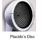
Shape of cornea is like Prolate spheroid. It is More curved in centre than periphery. Cornea is Transparent , Due to regular stromal collagen and presence of Na+-K+ ATPase pump in endothelium layer.90% light transmission occurs through cornea. Keratometer measures the cornea curvature and the technique is known as keratometry. Instruments used Placido’s disc-In normal cornea, there is no distortion of reflected pattern . In Keratoconus, distortion of reflected pattern is present
6 layers of cornea are:
- Epithelium
- Bowman’s membrane -Injury to this layer leads to corneal opacity or scar
- Stroma - it is the Thickest layer
- Predescemet’s layer (Dua’s layer)
- Descemet’s membrane -Strongest layer
- Endothelium -Single layer of hexagonal layer and it Maintain corneal opacity
Thickness of Cornea
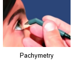
It is Thinnest in centre & Thickest at periphery, Ranging from 500 -600 μ, It is the Most powerful refracting surface eye: 43D (70 %) Cornea is avascular. IOP depends upon central corneal thickness (CCT). It is Measured by Pachymetry
Limbus
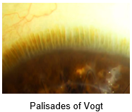
It is Junction of cornea and sclera Surgical limbus is zone of 2 mm . Corneal stem cells are present at limbus. It consists of Longitudinal parallel cells . it is Yellowish brown in colour . It is so known as Palisades of Vogt
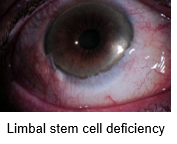
In case of deficiency of stem cells due to chemical injury (mc), prolonged contact lens wearing etc. there is decreased regeneration capacity of eye.
Signs of Limbal stem cell deficiency :
- Cells breakdown
- Inflammation
- Persistent epithelial defects
- Ulceration
- Neovascularization
- Conjunctivalisation
On Impression Cytology: Presence of Goblet cells on cornea (normally goblets cells are present in conjunctiva not in cornea)
Also Read: Pterygium: Causes, Symptoms, Diagnosis, and Treatment
MIDDLE LAYER OF EYEBALL
Uvea
Consists of
- Iris
- Ciliary Body
- Choroid
Iris
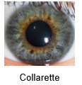
It is the Most anterior part of the uveal tract. It is Divided by collarette into central pupillary zone and peripheral ciliary zone two muscles of iris are Sphincter pupillae and Dilator pupillae.
Function - It Controls entry of light by dilating / constricting pupil . There are Two arterial circles :
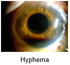
Major arterial circle - It Lies at root of iris where it attaches to the ciliary body and If it bleeds leads to Hyphema
Minor arterial circle - It Lies at collarette
CILIARY BODY
It has 3 parts :
- Pars Plicata: Anterior half with folds
- Pars Plana: Posterior half without folds
- Ciliary Muscle: Enclosed by Pars Plicate & Pars Plana
Root of the iris is attached to the ciliary body. It is the Thinnest part of iris.its Rupture leads to iridodialysis. Characteristic D-shaped pupils are seen in Iridodialysis. Its function is
Aqueous humour production, Accommodation. Pars plana only way to access vitreous humour Maximum risk of sympathetic ophthalmitis occurs on injury to ciliary body.
CHOROID
It is the Posterior most structure of uvea. It is Most vascular structure of eye. Choroid circulation gives 85% of ocular blood flow. Vortex Veins Drains entire uvea but particularly choroid)
Function - It Supplies nutrients to outer retina, and helps in Thermoregulation
4 layers of choroid are:
- Chorio-capillaries layer
- Bruch’s membrane
- Haller’s layer
- Sattler’s layer
Ophthalmology Related articles:
INNER MOST LAYER OF EYEBALL
Retina
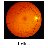
It is the Innermost layer stretches from central fovea to peripheral ora serrata which is Present only posteriorly. Central retina consists of Macula, fovea, optic disc Centre of retina occupied by Macula Centre of Macula Occupied by Fovea Fovea is most sensitive structure to light so brightest and sharpest image forms here.
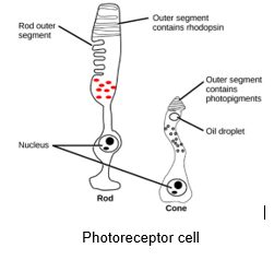
Microscopic Structure of Retina
It has Neurons of 5 Types .Neuroglia [supporting cells] of 3 Types
Neuron Types
- Photo Receptors (Rods/Cones) (1st Order neuron)
- Bipolar Cells (2nd Order neuron)
- Ganglion Cells (3rd Order neuron)
- Amacrine Cells
- Horizontal Cells
Neuroglia cells
- M - Muller’s
- A – Astroglia
- M – Microglia
Photoreceptors
|
Rods |
Cones |
|
|
This is everything that you need to know about layers of eye for your ophthalmology preparation.. For more interesting and informative blog posts like this download the PrepLadder App and keep reading our blog!

PrepLadder Medical
Get access to all the essential resources required to ace your medical exam Preparation. Stay updated with the latest news and developments in the medical exam, improve your Medical Exam preparation, and turn your dreams into a reality!
Navigate Quickly
Three Layers of Eye are
Outermost Fibrous Layer
Cornea
Thickness of Cornea
MIDDLE LAYER OF EYEBALL
INNER MOST LAYER OF EYEBALL
Microscopic Structure of Retina
Photoreceptors