Thanatology: Types of Death and Post Mortem Changes
Feb 13, 2026

The study of death and dying from a variety of reasons, including medical, physical, psychological, spiritual, ethical, and more, is known as Thanatology.
Death comes under section 46 of the Indian Penal Code(IPC).
Taphonomy is the study of post-mortem changes.
As we are studying thanatology, it becomes important for us to study the Components of Bishop's tripod of life. It includes the Heart(circulation), lung (respiration), and the brain function.
Thanatology is used by experts from a wide range of professions, from doctors and coroners to hospice workers and grief counselors, to inform their work. Additionally, there are Thanatology experts who concentrate on a particular component of the dying process or work directly with those who are confronting their own death or the death of a loved one.

Types of Death
Somatic death or Clinical death
In this, there is a complete and irreversible stoppage of any of Bishop’s tripod of life components, which includes
- Heart – circulation
- Lung – respiration
- Brain function
Or there is a loss of any one function, and it leads to death, or the Doctor declares the individual dead
Molecular death or Cellular death
It occurs within 1-2 hours after somatic death
Bichat’s mode of death
It depends on the dysfunction, i.e., it may be respiratory, circulatory, or brain function.
- Dysfunction of respiration leads to Asphyxia
- Dysfunction of circulation leads to Syncope
- Dysfunction of brain function leads to Coma
Asphyxia, syncope, and coma are examples of Bichat’s mode of death.
Atria mortis or Gateway of death
Stopping of only one component of Bichat’s tripod of life (respiration, circulation, or brain function) results in death. It is an example of somatic death.
Suspended Animation or Apparent Death.
In this, signs of life are reduced to a minimal level, and resuscitation leads to survival. The causes of suspended animation can be Prolonged anesthesia, Newborn (most common), barbiturate poisoning, Cholera, Cachexia, Concussion, Drowning, Electrocution, Hypothermia, Hyperthermia, Sunstroke, Shock, Insanity, Trance (voluntarily suspended animation seen in yoga practitioners), Typhoid (enteric fever), and Morphine overdose. Causes of suspended animation are:
- Prolonged anesthesia
- Newborn (most common)
- Barbiturates poisoning
- Cholera
- Cachexia
- Concussion
- Drowning
- Electrocution
- Hypothermia
- Hyperthermia
- Sunstroke
- Shock
- Insanity
- Trance (voluntarily suspended animation seen in yoga practitioners)
- Typhoid (enteric fever)
- Morphine overdose
Grab 100 One-Liners in FMT – Download Free PDF!
Post-Mortem Changes
Signs of death may be immediate, early, or late.
Early signs include:
- Eye changes
- Algor mortis
- Livor mortis
- Rigor mortis
Immediate signs include
- 1st sign is insensibility (loss of sensation) and loss of voluntary power
- There is a stoppage of respiration and circulation.
Late signs include:
- Decomposition signs
- Putrefaction
- Autolysis
- Mummification
Early Signs:
Eye changes
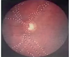
A) Railroading Phenomenon or Cattle Trucking or Kevorkian Sign
The Retinal blood vessels appear segmented or fragmented. It is seen within a few minutes to one hour after death. It can be seen using an ophthalmoscope. It is the earliest eye sign, and it is used to determine time since death (TSD). Before the Kervokian sign, corneal reflexes are lost, and the pupil is dilated.
The "Kevorkian sign is the fragmentation or trucking of blood vessels, and it is a crucial ophthalmoscopic finding. Within a few minutes of death, the Kevorkian sign appears and lasts for around an hour.
Kevorkian sign
B) Flaccidity of the Eyeball
Normal intraocular pressure is 20 mmHg. Two hours after death, it drops to 0 mmHg. It is also used to determine the time since death.
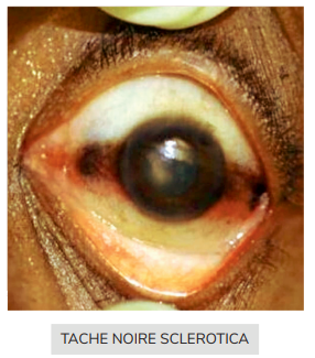
c) Tâche Noire Sclérotique
After death, if the eyelids are open and dust is present in the atmosphere, it will get deposited in the sclera, resulting in the formation of 2 brownish triangular-shaped opacities on either side of the cornea, which is known as Tache Noire Sclerotique. It occurs after 3-6 hours of post-mortem and is used to determine time since death.
D) Corneal changes
Normally, the cornea is transparent, but after 1 hour of death, it becomes hazy. Conversely, opacity occurs after 6 hours.

Vitreous Humor Changes
It is the most important medium in the eye that resists putrefaction. There are no chemical changes in the vitreous humor after putrefaction. It is the best medium for estimating time since death, as it is enclosed within the eyeball, so bacteria cannot have access to it easily.
So, even in an advanced decomposed body, this medium is very helpful. The best parameter is vitreous potassium levels. There is a linear increase in K+ and hypoxanthine. It has a linear correlation with time since death. Two formulas used for finding time since death are Sturner's Formula and Madea's Formula.
Also Read: Pectus Excavatum: Causes, Symptoms, Risk Factors, Diagnosis and Treatment
Cooling of the body or Algor mortis
It is seen within 15 minutes after death. In this, the body core temperature (BCT) decreases. A thanatometer is a chemical thermometer used to measure BCT in post-mortem. It may be 25-30 cm. It can measure temperatures between 0-50°C. The ideal site for recording temperature is the rectum (rectal temperature is almost equal to BCT).
Other sites include the inferior surface of the liver, the external auditory meatus, the nasal spaces, and the lower end of the esophagus. Graph illustrating the relationship between temperature and time post-mortem produces a sigmoid curve or inverted S-shape curve.
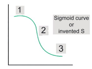
- Stage I: Isothermic phase.
- Stage II: Steep decline.
- Stage III: Gradual decrease.
Algor mortis occurs in 3 phases
- 1st phase: Gradual decrease in BCT
- 2nd phase: Rapid decrease in BCT
- 3rd phase: Gradual decrease in BCT · Shape of Algor mortis curve is Sigmoid.
The rate of temperature fall is 0.4°C —0.7°C / hr. In summer, it is 0.4° C / hr, and in winter, it is 0.7° C / hr. The average fall in temperature is 0.5 C/hr. Knowing the rate of fall, we can calculate the time since death.
Post-Mortem Caloricity
Normally, the body becomes cold within 15 minutes after death [Algor mortis]· But if the body remains warm for 1-2 hours even after death, it is known as Post-mortem caloricity. Whenever there is an increased body core temperature at the time of death, post-mortem caloricity is seen.
Increased body core temperature at death is seen in conditions with increased Muscle contraction, that is, Tetanus, Strychnos Nux vomica poisoning, exercises, etc. Defective thermoregulation in the body is associated with Heat stroke, Pontine hemorrhage [have pyrexia, paralysis, pinpoint pupils], and Septicemia.
Post-Mortem Staining
It is also known as post-mortem lividity, hypostasis, livor mortis, vibices, or suggillation. This stain is Present in dependent parts of the body, i.e., areas that do not touch the ground but face towards the ground.

Gravitational forces cause blood to pool in capillaries and venules, accumulating deoxyhemoglobin blood in the skin, and causing a bluish-purple appearance. There is also Skin discoloration of the rete mucosum of the dermis.
This stain is absent in the pressure parts of the body. It begins shortly after death, that is, after 30 minutes. And it is visible for 2 hours after death. Maximum visibility is for 6-12 hours, and fixation occurs after 7-8 hours.
Secondary lividity occurs when changing the position of the body before 7-8 hours, leading to post-mortem staining in other areas of the body, which persists till decomposition. Decomposition changes the skin color to green.
Post-mortem staining in different positions
- In the Supine position → the back of the head, back of the neck, back of the chest, and back of the leg are stained
- In the prone position → the front parts of the body are strained.
- In the Hanging position → the hands and feet are stained, i.e., show glove and stocking pattern.
- In Running water → no post-mortem staining is seen
Medical importance
It helps to determine the time since death, the position of the body at the time of death, and the cause of death (COD).
|
|
|
|
|
|
|
|
|
|
|
|
Color Presentation due to Different Causes of Poisoning:
- Carbon monoxide poisoning → Cherry-red
- Cyanide poisoning → Bright or brick red
- Hypothermia → Pink
- Hydrogen sulfide (H2S) → Blue green
- Opium → Black
- Phosphorus or potassium chlorate (KCLO3) → Chocolate brown.
- Nitrites, nitrobenzene, aniline → Chocolate brown
- Aniline may produce a blue color.
- The chocolate brown color formed due to methemoglobin.
- Clostridium perfringens → Bronze
- Methanol poisoning → Purple
Rigor Mortis or Cadaveric Stiffening or Cadaveric Rigidity
It is also known as cadaveric rigidity / Cadaveric stiffening. It gives the muscle status after the death of a person. The decrease in ATP causes stiffening of the muscle. Rigor mortis is generalized, that is, it is seen in both voluntary & involuntary muscles, but it is first seen in involuntary muscles.

3 stages muscles undergo after death include:
- Primary flaccidity phase or primary relaxation phase.
- Rigor mortis.
- Secondary flaccidity phase or secondary relaxation phase.
Primary flaccidity occurs during somatic death. Rigor mortis occurs during cellular death. The mechanism of rigor mortis is ATP depletion. Actin-myosin separation takes place in primary flaccidity. Lack of actin-myosin separation causes rigor mortis.
Secondary flaccidity occurs when decomposition starts in this Actin and myosin filaments are broken down, leading to separation. When rigor mortis starts, ATP decreases to 85% of the normal content. At maximum rigor mortis, ATP decreases to 15-30% of the normal content.
Occurrence of Rigor Mortis in Muscles
It begins 1-2 hours after death. It is seen in all muscles, whether voluntary or involuntary. 1st muscle affected is the myocardium. 1st external muscle affected is the upper eyelid muscle, i.e., orbicularis oculi. Sequence in which external muscles are affected:
Eyelid muscles → Neck → Lower jaw → Muscles of face → Muscles of chest → Upper limbs → Abdomen → Lower limbs → Fingers and toes
Rigor mortis disappears in the same order that it appears. It will disappear first from the upper eyelid and last from the fingers and toes.
Rule of 12
As we discuss thanatology, it becomes very important to mention the Rule of 12 in forensic medicine.
- In the first 12 hours after death, rigor mortis appears in all body muscles.
- In the next 12 hours, it persists in all body muscles.
- In the next 12 hours, it disappears from all body muscles.
- Rigor mortis appears and disappears within 36-48 hours after death, thereby aiding in the determination of TSD.
Rigor Mortis in a Fetus
Rigor mortis is absent in fetuses less than 7 months of age because the Actin and myosin filaments have not developed. Rigor mortis' appearance varies according to the season. In summer, it is seen in 18-36 hours, whereas in winter, it is seen after 24-48 hours.
Rigor Mortis Appears in Wasting Diseases
This causes muscle mass to be thin with decreased ATP storage. It is seen in Diseases such as cholera, TB, cancer, cachexia, and typhoid. There is also an Early onset of rigor mortis with a short duration.
Rigor Mortis appears in Violent Death
In Cases such as cutthroat injury or firearm injury. It has an early onset of rigor mortis with a short duration. It is important to remember two exceptions here:
In Strychnine poisoning, there is an Early-onset rigor mortis with long duration.
In Arsenic poisoning, we will have a late onset of rigor mortis with a long duration.
Rigor Mortis Appearance in Thick Muscle
There is increased ATP storage, and it has a late onset of rigor mortis.
Related Articles:
Causes of Muscle Stiffening after Death
- Rigor mortis
- Heat stiffening
- Cold stiffening
- Gas stiffening
- Cadaveric spasm
Heat Stiffening
Heat stiffening is also known as the boxing attitude, pugilistic attitude, or fencing attitude. It occurs when the Surrounding external temperature is ≥ 65°C. The mechanism involves the coagulation of muscle protein. Normal rigor mortis is not seen. Heat stiffening persists until decomposition occurs.
Cold Stiffening
Cold stiffening occurs when the surrounding external temperature is ≤ -5°C. The mechanism involves freezing of the body fluid and hardening of the subcutaneous tissue. In warm temperatures, cold stiffening disappears. In this, Normal rigor is present.
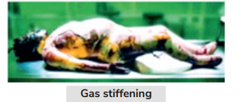
Gas Stiffening
Gas stiffening is seen with decomposition. During decomposition, excess gas is produced. These gases accumulate in the body, resulting in stiffness.
Cadaveric Spasm
It is also known as cataleptic rigidity or instantaneous rigor. The mechanism involves ATP depletion. Muscles remain contracted for a lifetime and remain rigid and stiff after death. It occurs immediately after death. Primary relaxation is absent, whereas Secondary relaxation is present. It is an exclusively ante-mortem event.

Causes of cadaveric spasm:
- Asphyxial death
- Brain injury
- Cerebral injury
- Drowning
- Dinitrocresol poisoning
- Excitement
- Fear
- Firearm, e.g., in suicide
Differences between cadaveric spasm and rigor mortis
Mainly involves voluntary muscles.
Generally, it involves a short group of muscles, e.g. hand muscles.Cadaveric spasm Rigor mortis Time Immediately after death. 1-2 hours after death. Muscles Mainly involves voluntary muscles.
Generally, it involves a short group of muscles e.g. hand muscles.Involves both voluntary and involuntary muscles. Primary relaxation Absent. Present. Molecular death Absent. Present. Electronic stimuli Response present. Response absent. Importance Describes the manner of death. Determines TSD.
Late changes
In these changes, the body decomposes. Decomposition occurs by two mechanisms: autolysis and putrefaction.
Autolysis
It is a process in which Body enzymes cause cell lysis. Examples of autolytic changes:
- Clouding of the cornea – 1st external change.
- Changes in brain glandular tissue – 1st internal change.
Aseptic autolysis involves the mummification of the fetus in intrauterine death.
Putrefaction
It is stimulated by bacterial activity. The most common bacteria involved are Clostridium welchii or Clostridium perfringens. The most common enzyme involved is lytic lecithinase.
Putrefaction occurs in three stages of change:
Putrefaction occurs in three stages of change:
- Color change
- Gas formation
- Liquefaction of tissue
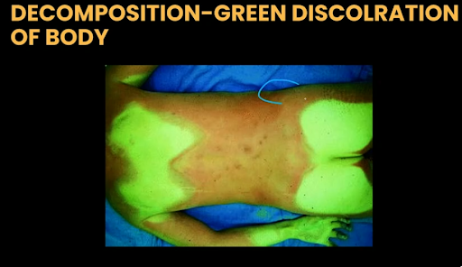
Color change:
1st internal/overall change of putrefaction is reddish-brown discoloration of the aortic intima.
1st external change is green discoloration of the right iliac fossa.
In summer, discoloration starts in 12-18 hours; in winter, discoloration starts in 1-2 days.
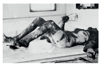
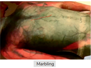
Marbling is the Discoloration of prominent superficial veins to green-brown color, which corresponds to the vascular channel. It starts in 24 hours and is prominent in 36-48 hours. It helps in determining Time since death (TSD). It is seen in the chest, shoulder, abdomen, and thigh.
Liquefaction Of Tissues
It takes 5-10 days. Loosening of hair/ nails occurs in 3-5 days, and skin around the hands & legs peels off, i.e. Degloving/Destocking pattern is seen in 5-10 days
Post-mortem purge
- Gas formation during decomposition leads to the expulsion of blood-stained froth and gastric contents from the nose and mouth.
- Seen after 2-3 days.
Post-mortem luminescence
The body emits light after death. Causes of post-mortem luminescence are:
- Bacteria, e.g., photobacteria, are present.
Fungal, e.g., Armillaria, Ram’s bottom.
Casper’s Dictum
It is about the rate of putrefaction in different media. The rate of putrefaction is compared b/w 3 imp medium. This Formula was proposed by Taylor. It shows the relationship between the rate of decomposition in air, water, and soil (earth).
- Air > Water > Earth.
If decomposition in the air takes 1 week, it will take 2 weeks in water and 8 weeks in soil. Air has the fastest rate of decomposition.
Putrefaction sequence
- 1st organs to decompose are the larynx and trachea due to their direct contact with air.
- Followed by (in order):
- Stomach, spleen, and small intestine.
- Liver and lung.
- Brain, heart, kidney, uterus.
- Skin, muscle, tendon, bone/tooth.
- The non-gravid uterus is the last organ to decompose in females.
- The prostate is the last organ to decompose in males.
- The overall last organ to decompose is bone/tooth.
Important points of putrefaction
- Liver shows gas formation up to 24-36 hours, i.e., foamy liver or honeycomb liver.
- The optimum temperature for decomposition is 21-38°C.
- Decomposition starts at > 10°C.
- Poisons inhibiting putrefaction include:
- Strychnine.
- Metallic poison, e.g, arsenic, antimony, thallium.
- Carbon monoxide.
- Cyanide.
- Carbolic acid.
- The 1st amino acid that disappears from bone in decomposition is proline.
- The 2nd amino acid to disappear is hydroxyproline.
- The last amino acid to disappear is glycine.
- Bones that are 100 years old have < 7 amino acids.
Modification of Putrefaction
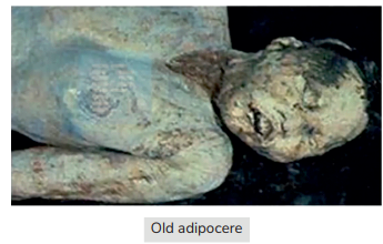
Adipocere or saponification or grave wax: The Fat in the body gets converted into fatty acids and combines with Calcium in the body & forms insoluble wax-like substances, i.e., (SOAP) adipocere.
Appearance of adipocere :
Fresh adipocere looks greasy, like rancid butter. Old adipocere looks dry, hard, yellow, and brittle. The time required for adipocere formation is 3 days to 3 months, and it is Absent in fetuses of less than 7 months.
Medical importance
It helps to describe the climate at the time of death and Determination of Time since death, and helps in the preservation of the dead body for easy identification.
Mummification
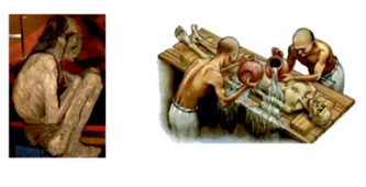
It has no specific smell/odor. It takes 3 months to 12 months to occur. Mummification is also seen in the intrauterine death of the fetus when there are intact membranes & deficient blood supply · Arsenic & Antimony favor mummification. It is an important feature of intrauterine death.
The Medical importance of mummification is that, other than describing the climate at the time of death and the time since death TSD, mummification helps us to identify the body comparatively very easily.
Embalming or Tanatopraxia is the Artificial method of preserving the body by injecting antiseptics & preservatives. The ideal time for embalming is less than 6 hrs after death (Very effective).
The preservatives of embalming fluid are formalin, formaldehyde, and methanol. The germicide in the embalming fluid is phenol, and the anticoagulant used is sodium citrate.
Ethanol is not a part of embalming fluid.
.jpg)
Ante-mortem vs Post-mortem
Ante-mortem Post-mortem Content Inflammation.
Chloride.
Protein (mainly, albumin).Gas. Base Erythematous. Pale. Redness Present. Absent. Enzymatic/Vital reaction Present. Absent.
Maggots
- Larvae of fly: Musca domestica/ vicinia
- Life cycle
- Summer: 5-10 days
- Winter: 8-20 days
- Lay eggs: Natural orifices
- Developmental stages
- Maggots are seen in 1-2 days
- Pupae: 3-6 days
- Adult flies : 3-6 days
- Rainy season: Maggots come earlier
- Determines TSD

Marbling Vs Filigree Burns
CRITERIA MARBLING FILIGREE BURNS Color Green- Brown Pink and erythematous Areas Shoulder, chest, neck, thigh, abdomen Correspond to vascular channels Yes No Mechanism Bacteria → H2S gas → H2S +Hb →Sulfhemoglobin (SulfHb) Superficial veins: Stain/prominent Denaturation of RBCs(Hb-Stain) Linear branching Lightning injury Starts in 24 hours → Prominent in36-48 hoursDetermines TSD

Important Mcq's
Q. Which of the following is false about the Kevorkian sign?
- Segmentation of the blood column in the carotid vessels
- Seen within minutes after death
- Seen by ophthalmoscope
- Little practical value
Ans: 1) Segmentation of the blood column in the carotid vessels
Q. Postmortem lividity persists till?
- 6 hours
- 24 hours
- 36 hours
- Putrefaction sets in
Ans: 4) Putrefaction sets in
Q. Which of the following eye signs can be appreciated in the image?

- Tache noir
- Kevorkian sign
- Corneal changes
- Vitreous humour changes
Ans: 1) Tache noir
Q. Identify the postmortem change given in the following image:

- Marbling
- Tattooing
- Lividity
- Filigree burns
Ans: 1) Marbling
Q. Identify the odor produced by the change seen in the image below:

- Odorless
- Pungent
- Putrid
- Offensive
Ans: 1) Odorless
Q. Postmortem luminescence is produced by which Bacteria/micro-organisms?
- Photobacterium fischeri
- Armillaria mellea
- Clostridium welchii
- Both A and B
Ans: 4) Both A and B
Q. Hypostasis is developed in which layer of the skin?
- Epidermis
- Dermis
- Hypodermis
- Just below the skin
Ans: 4) Just below the skin
Q. Earliest internal sign of decomposition is:
- Greenish discoloration under the liver
- Liquefaction of the pancreas
- Reddish-brown discoloration of the inner surface of the aorta
- Congestion of the spleen
Ans: 3) Reddish-brown discoloration of the inner surface of the aorta
Q. First organ to undergo putrefaction is:
- Larynx
- Liver
- Stomach & intestine
- Prostate
Ans: 1) Larynx
Q. Which of the following is false about cadaveric spasm?
- Molecular death does not occur
- It involves a single group of muscles
- Muscle passes through a stage of primary relaxation
- Helps in deciding the manner of death
Ans: 3) Muscle passes through a stage of primary relaxation
Q. The bluish-purple color of the postmortem lividity is due to:
- Oxy hemoglobin
- Deoxy-haemoglobin
- Met-haemoglobin
- Sulphmethaemoglobin
Ans: 4) Sulphmethaemoglobin
If you're looking to test your preparation and gear up for the NEET PG exam, the NEET PG Mock exam is always available for you. This comprehensive mock exam, designed to mirror the actual exam pattern and difficulty, is available throughout your preparation to assess your progress.
Download the PrepLadder app now and unlock a 24-hour FREE trial of premium high-yield content. Access Video Lectures, digital notes, QBank, and Mock Tests for FREE to ace your NEET PG preparation. Elevate your study experience and gear up for success. Start your journey with PrepLadder today!

PrepLadder Medical
Get access to all the essential resources required to ace your medical exam Preparation. Stay updated with the latest news and developments in the medical exam, improve your Medical Exam preparation, and turn your dreams into a reality!
Navigate Quickly
Types of Death
Somatic death or Clinical death
Molecular death or Cellular death
Bichat’s mode of death
Atria mortis or Gateway of death
Suspended Animation or Apparent Death.
Grab 100 One-Liners in FMT – Download Free PDF!
Post-Mortem Changes
Early Signs:
Cooling of the body or Algor mortis
Post-Mortem Staining
Rigor Mortis or Cadaveric Stiffening or Cadaveric Rigidity
Causes of Muscle Stiffening after Death
Differences between cadaveric spasm and rigor mortis
Late changes
Mummification
Ante-mortem vs Post-mortem
Maggots
Marbling Vs Filigree Burns
Important Mcq's
Top searching words
The most popular search terms used by aspirants
- Forensic Medicine Preparation
- NEET PG FMT
- NEET PG FMT Preparation