NEET PG/FMGE Ophthalmology Important Questions With Answers
Dec 5, 2024

Ophthalmology is undeniably one of the most critical yet challenging subjects in FMGE syllabus. You must have a clear understanding of clinical applications and not just theoretical knowledge.
From diagnosis of complex conditions and retinal disorders to intricate anatomy of the eye, every minute detail matters when you’re preparing for such a high-stakes exam.
We have curated a comprehensive list of high-yield questions and answers on Ophthalmology to help you navigate this vast subject. These questions are sure to reinforce your understanding and also boost your confidence to tackle the exam head-on.
Read the blog further to gain clarity on the most frequently asked topics and streamline your FMGE preparation.
Q. What are the number of ciliary processes?
Ans. There are 70-75 ciliary processes in a single eye.
Q. What is the normal ocular pressure?
Ans. The normal ocular pressure is 10-21 mmHg.
Q. When the pressure is more, what happens?
Ans. When the ocular pressure is higher, it damages the optic nerve. This condition is known as glaucoma.
Also read: NEET PG High-Yield Questions on Parasitology
Q. What is lamina cribrosa?
Ans. When the optic nerve exits through the eye, and the sclera at the disc is perforated, known as lamina cribrosa.
Q. The nerve fibre of the retina that aggregates at the disc to form.
Ans. Optic nerve
Q. What is the capacity of the orbit?
Ans. It is 30 cc. The orbit is quadrilateral and pyramidal in shape.
Q. What is the axial length of the eye?
Ans. It is around 24 mm.
Q. What is the depth of the anterior chamber?
Ans. 2.4-2.5 mm. 2.5 comes as the definition of anisometropia. Infants are also hypermetropic by 2.5-3 D.
Also read: Important Psychiatry One-Liners for NEET PG/FMGE
Q. How is the axial length measured?
Ans. Axial length is measured by an A-scan.
Q. Total refractive power of the eye is?
Ans. 58-60 D
Q. Total refractive power of the cornea is?
Ans. 43-45 D
Q. Total refractive power of the lens is?
Ans. 16-17 D
Q. What is the index of the cornea?
Ans. 1.37
Also read: Ocular Rosacea: Causes, Symptoms, Diagnosis, Treatment
Q. What is the index of the lens?
Ans. 1.39. It is maximum at the centre of the lens (1.4-1.41).
Q. Which is the thinnest part of the lens?
Ans. The capsule at the posterior pole
Q. Till when are lens fibres formed?
Ans. They are formed throughout life
Q. Which are the youngest fibres?
Ans. Cortexes are the youngest fibres
Q. Where is the lens derived from?
Ans. Surface ectoderm
Also read: Melanoma Of Eye: Causes, Symptoms and Risk Factors
Q. What is the primary metabolism of the lens?
Ans. It is anaerobic. 80% of glucose is metabolised anaerobically.
Q. How to differentiate pterygium from pseudopterygium?
Ans.
- Glass Rod Test: The glass rod test will not pass through the pterygium.
- In pseudopterygium, it will pass.
Q. Where are the maximum goblet cells found?
Ans. Goblet cells are found most frequently in the inferonasal quadrant.
Q. Nasolacrimal gland opens into?
Ans. Inferior meatus of the nose.
Q. Where is the inferior meatus present?
Ans. Anteriorly
Also read: Eye Strain: Causes, Symptoms, Risk Factors, Diagnosis
Q. What is the alignment of the upper and lower puncta?
Ans. Upper puncta is medial to the lower puncta.
Q. What is the treatment strategy to diagnose the lacrimal pump failure?
Ans. Dacryoscintigraphy
Q. What is the treatment strategy to diagnose the lacrimal drainage system?
Ans. John Titus
Q. Where is the opening made in DCR?
Ans. Medial meatus
Q. What is the direction of the probe?
Ans. In the nose's direction (downward, more laterally, and backward).
Also read: Subconjunctival Haemorrhage: Causes, Symptoms, Risk Factors
Q. What is the treatment of lacrimal fistula?
Ans. Dacryocystorhinostomy (DCR)
Q. Which is the most common organism for the pathogenesis of lacrimal fistula?
Ans. Staphylococcus aureus
Q. What is the treatment for chronic dyseryocystitis?
Ans. Dacryocystorhinostomy (DCR)
Q. Is chronic dyseryocystitis more common in?
Ans. Females
Q. The most common organism for pathogenesis is?
Ans. Staphylococcus aureus
Also read: Fuchs' Dystrophy: Causes, Symptoms, Risk Factors, Diagnosis and Treatment
Q. What is the treatment of choice for lacrimal fibrosis?
Ans. Dacryocystectomy
Q. Why are keratin proteins always present at the lower cornea?
Ans. Due to the convection current of aqueous humour.
Q. What are the signs of active inflammation?
Ans. Aqueous cells
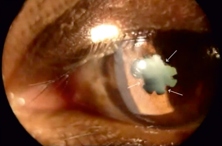
Q. The point where these aqueous cells adhere or not move?
Ans. Festoon shape pupil
Also read: Optic Neuritis: Causes, Symptoms, Risk Factors, Diagnosis and Treatment
Q. What is the treatment of choice for anterior uveitis?
Ans.
- Topical steroids and Topical cycloplegic
- Topical cycloplegic relaxes the muscles by relieving the ciliary spasm and prevents posterior synechiae
Q. What is the Luminate program?
Ans. The Luminate program assesses the safety and efficacy of voclosporin for the treatment of all forms of uveitis.
Q. Identify the picture?
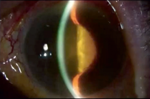
Ans. Iris Bombe
Leads to angle closure
Q. What is the complication of FHI?
Ans. Complicated cataract (particularly posterior subcapsular cataract) and secondary glaucoma
Also read: Blindness: Causes, Symptoms, Types, Diagnosis, Treatment and Prevention
Q. What is the characteristic feature of CMV retinitis?
Ans. CMV retinitis causes haemorrhage and necrosis.
Q. What is the most common opportunistic infection in HIV?
Ans. CMV retinitis
Q. What is the most common feature of HIV?
Ans. Microangiopathy, microscopic examination reveals cotton wool spots.
Q. Headlight in fog appearance
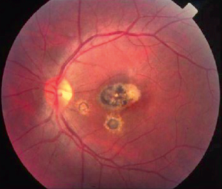
Ans. Foggy lesions (intense vitritis); lesions look like fog, which is the characteristic feature of toxoplasmosis and the most common
cause of anterior or posterior uveitis. When it heals, it leads to punched-out pigmented lesions, especially in the macula area, and vision goes down.
Q. What is the time duration of SO?
Ans. Never before 2 weeks because it is an autoimmune reaction, and it will take time to occur.
Also read: Retinitis Pigmentosa: Causes, Symptoms, Diagnosis and Treatment
Q. What type of uveitis is this?
Ans. Granulomatous Pan Uveitis.
Q. Where are the nodules present?
Ans. These nodules are present between Bruch's membrane and retina.
Q. What is the first sign of SO?
Ans. A retrolental flare is the first sign; difficulty in near vision is the first symptom.
Q. What is the length of the optic nerve?
Ans. 3.5 to 5 cm
Q. Which is the longest part of the optic nerve?
Ans. Intraorbital part
Q. What is the length of the intraorbital part?
Ans. 25-30 mm long
Also read: Keratoplasty: Procedure, Results, Complications For Keratoplasty
Q. What is the first sign of optic nerve disease?
Ans. Relative afferent pupillary defect (RAPD)
Q. What can be the field effects when the nerve is involved?
Ans. Central scotoma, When the scotoma joins the binding spot and macula, it is called the centrocecal scotoma.
Q. What is the main cause of centrocecal scotoma?
Ans. Tobacco, due to the presence of cyanide in it, destroys the optic nerve and is called tobacco amblyopia.
Q. Nerve fibre defect (Bjerrum Scotoma) can be seen in?
Ans. Glaucoma
Q. Altitudinal defect is the key feature of?
Ans. Anti-ischemic optic neuropathy (AION)
Also read: Corneal Ulcer: Causes, Symptoms, Risk Factors and Diagnosis
Q. How does the patient present with an optic nerve disorder?
Ans.
- Decreased visual acuity.
- Visual field defect.
- Relative afferent pupillary defect (first sign)
- Decrease in a red-green colour called dyschromatopsia (exception: glaucoma).
- Decrease in brightness.
Q. What is a total afferent pupillary defect?
Ans. When there is total atrophy and no reaction at all, which is the feature of optic atrophy.
Q. Which cells are responsible for colour vision?
Ans. Parvocellular cells.
Q. Where is the keyhole visual defect seen?
Ans. LGB lesion
Q. Defective Optokinetic Nystagmus (DON) is seen in?
Ans. Deep parietal lobe lesion.
Also read: Retinoblastoma: Causes, Symptoms, Diagnosis and Treatment
Q. What is the capacity of the orbit?
Ans. 30cc
Q. What is the shape of the orbit?
Ans. Quadrilateral or pyramidal in shape.
Q. Which is the weakest wall of the orbit?
Ans. Medial wall because of the presence of lamina propria.
Q. Which is the weakest wall of the floor?
Ans. Posteromedial part.
Q. Any fracture in the orbit is called?
Ans. Blow out fracture.
Q. Which instrument is used for the diagnosis of proptosis?
Ans. Hertel's exophthalmometer.
Also read: Refractive Errors: Types, Diagnosis, Symptoms and Treatment
Q. Which instrument is used in children for the diagnosis?
Ans. Luedde ophthalmometer
Q. Which is the first muscle to be involved?
Ans. The first inferior rectus muscle and the last muscle, the inferior oblique muscle, is involved.
Q. Which is the first defective movement?
Ans. Defective elevation because of the fibrosis of the inferior rectus muscle.
Q. Which test is used to diagnose palsy and fibrosis?
Ans. In the forced duction test, holding the superior rectus muscle, tries to move the eye up. If with the forceps it moves easily, then it is a case of palsy; Even with the forceps it is stuck in one position, then it is fibrosis (positive)
Q. What is the first sign?
Ans. Lid sign, which is retraction.
Also read: Blinking Of Eye: Types, Uses, Causes, Diagnosis and Treatment
Q. Why is there a need for an ocular emergency or why the patient needs to be admitted?
Ans. Because there is a risk of cavernous sinus thrombosis (CST).
Q. What is the first sign of cavernous sinus thrombosis (CST)?
Ans. VI nerve palsy. The 3,4,5 nerve passes the wall of the sinus, and the 6th nerve passes through the body along with the internal carotid artery. Any granulomatous idiopathic inflammation of the cavernous sinus along with superior orbital fissure and orbital apex is called Tolosa Hunt syndrome. The patient is admitted, and treatment starts with I.V. antibiotics (both aerobic and anaerobic), then I.V. anti-inflammatory.
Q. What is the aetiology of orbital cellulitis?
Ans. Streptococcus, Staphylococcus aureus, and in children, it is Haemophilus.
Q. What is the most common type of lacrimal gland tumour?
Ans. Pleomorphic adenoma
Also read: Dacryocystitis : Causes, Types, Symptoms, Diagnosis
Q. What is the most common type of Malignancy?
Ans. Adenoid cystic carcinoma
Q. Which is the most dangerous malignancy?
Ans. Adenoid cystic carcinoma because of perineural invasion.
Q. Which is the most common intraorbital tumor of adults?
Ans. Cavernous haemangioma, Cavernous haemangioma is encapsulated, intraconal, and leads to axial proptosis.
Q. Optic nerve glioma is more common in?
Ans. Patients with NF-1
Q. Which is the most common pathology?
Ans. Pilocytic astrocytoma
Q. What is the finding of fusiform enlargement on CT-scan and MRI?
Ans. Fusiform enlargement of the optic nerve
Also read: Coat Disease: Causes, Symptoms, Stages, Diagnosis and Treatment
Q. What is the most common ocular feature of NF-2?
Ans. Posterior subcapsular cataract
Q. Which is the most common muscle involved?
Ans. Inferior rectus muscle/inferior oblique muscle
Q. Which is the most dangerous foreign body?
Ans. Copper that leads to phthisis bulbi. The most common mode of injury is chisel and hammer.
Q. How can siderosis be diagnosed?
Ans. Serial electroretinogram (ERG), mainly B-wave, is diminished.
Q. Any exposure keratopathy due to 7th nerve palsy called?
Ans. Neuroparalytic keratitis
Q. What is the most common feature of ptosis?
Ans. Aponeurotic
Also read: Ectropion: Causes, Symptoms, Risk Factors and Diagnosis
Q. What is the amount of dioptre that can be compensated by using the pinhole?
Ans. 3D
Q. What is the width of the stenopic slit?
Ans. 1 mm
Q. Which is the most common colour blindness?
Ans. Green blindness
Q. Discuss the Ishihara chart?
Ans. Ishihara chart is used for screening purposes (red-green blindness)
Q. Where is the image formed in an indirect ophthalmoscope?
Ans. Image formed between the examiner and lens.
Q. What type of image is formed?
Ans. Real/inverted magnification is around 3 or 5 times.
Also read: Entropion: Causes, Symptoms, Risk Factors and Diagnosis
Q. What is the field?
Ans. 8DD (8×1.5mm)
Q. How do you calculate the magnification of an indirect ophthalmoscope?
Ans. Power of eye/Power of lens
Q. Magnification is calculated by?
Ans. Power/4
Q. If magnification is more, then the field will be?
Ans. Less, which is around 2 DD
Q. If you examine the right eye of the patient, which eye will you use?
Ans. Always the same eye, and the patients are instructed to look straight.
Q. Is a direct ophthalmoscope generally used to see?
Ans. Central retina
Also read: Eye Floaters: Causes, Symptoms, Risk Factors, Diagnosis and Treatment
Q. What is a distance direct ophthalmoscope?
Ans. When it is done through a distance of 25 cm.
Q. Pin-Cushion effect due to?
Ans. Pin: High spherical aberration.
Q. What is the treatment of choice for aphakia?
Ans. Ideally, it is a PC IOL.
Q. With the near point of 20 cms, a hypermetrope of +4 has to exercise accommodation of:
- +11D
- +9D
- +1D
- None of the above
Ans. +9D
Q. What are the tests to check binocular single vision?
Ans. Worth four-dot test
Also read: Myopia: Causes, Symptoms, Risk Factors and Diagnosis
Q. What percentage deviation is there for infantile esotropia?
Ans. There is a large percentage of deviation, up to 30% D.
Q. How many months does infantile esotropia manifest?
Ans. It can manifest after 6 months.
Q. What is latent nystagmus?
Ans. When one eye is covered, there is nystagmus in the child's eye.
Q. What is dissociated vertical deviation (DVD)?
Ans. When the eye is covered, one eye goes up and out. It is a neurological defect. It does not obey Hering's law.
Also read: Important Psychiatry One-Liners for NEET PG/FMGE
Download the PrepLadder app now and unlock a 24-hour FREE trial of premium high-yield content. Access Video Lectures, digital notes, QBank, and Mock Tests for best neet pg ophthalmology online coaching. Elevate your study experience and gear up for success. Start your journey with PrepLadder today!

PrepLadder Medical
Get access to all the essential resources required to ace your medical exam Preparation. Stay updated with the latest news and developments in the medical exam, improve your Medical Exam preparation, and turn your dreams into a reality!
Top searching words
The most popular search terms used by aspirants
- NEET PG Ophthalmology
- NEET PG Ophthalmology Preparation