Intestinal Ulcers and Infections - NEET PG Pathology
Feb 17, 2023

Intestinal ulcers and infections are common conditions encountered in clinical practice, significantly impacting morbidity and mortality. Many other gastrointestinal conditions may present symptoms like intestinal ulcers and infections. And thus, it is important to have a thorough understanding of the etiology, pathophysiology, clinical features, and management of these conditions.
Read this blog further to get a quick overview of this important pathology topic for NEET PG exam preparations.

Tuberculosis

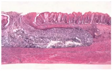
- The ulcers found in intestinal tuberculosis are transverse ulcers.
- Intestinal tuberculosis is seen secondary to the development of pulmonary tuberculosis.
- Pathogen: the pathogen associated with intestinal tuberculosis is Mycobacterium bovis.
| Important information Mycobacterium bovis, an organism that is present in contaminated dairy products, is also responsible for interstinal tuberculosis sometimes. The common route of infection is usually through the injection of contaminated sputum. The moment the transverse ulcer heals, it is going to be associated with fibrosis and leads to thickening of the bowel wall. The structure thus leads to a common surgical emergency called Subacute intestinal obstruction. |
Enteric fever/typhoid

- Pathogen associated with typhoid is Salmonella typhi.
- It is an infection that spreads through the intake of contaminated water and food.
- Typhoid fever is associated with the development of longitudinal ulcers.
- This kind of an ulceration is likely to be associated with bleeding, usually in the third week; but less likely to be associated with a stricture formation.
| Important information Erythrophagocytosis caused by the ingestion of RBCs by a macrophage is one of the most important findings that can be found in an individual who is having typhoid ulcer. |
Hints to look for
- Fecal-oral transmission
- History of involvement of abdominal pain and fever.
- Involvement of ileum. Because the ulcers are usually found at the level of the Peyer’s patches.
- Microscopically, there will be the presence of erythrophagocytosis.
Amoebiasis
- Spreads through the ingestion of contaminated water.
- CAUSATIVE ORGANISM: Entamoeba histolytica.
- LOCATION: intestine
Important information
- Since the ulcer is unable to enter the muscle layer, it takes a “flask-shape” (flask shaped ulcer formation)
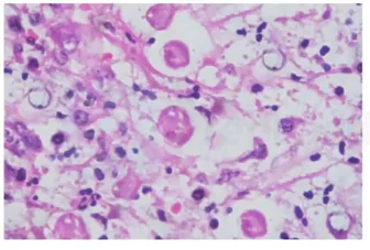
- MICROSCOPIC VIEW:
- The trophozoite form is going to have the presence of a round nuclei.
- Some of them might have ingested RBCs.
- The trophozoites can get access to the portal circulation and thereby reach the liver. In the liver, they can be responsible for the formation of amoebic liver abscess.
| Important information The key word to be noted regarding amoebic liver abscess is “anchovy sauce” pus. Antibiotics like metronidazole is used for the treatment of amoebic liver abscess. |
Also Read: Thalassemia - Symptoms and Causes - NEET PG Pathology
SICKLE CELL ANEMIA AND THALASSEMIA - NEET PG Pathology
CHRONIC INFLAMMATION: Symptoms, Causes, Diagnosis, and Treatment: Pathology
Amyloidosis: Types, Causes, Symptoms, Diagnosis, Risk Factors, Treatment

Psuedo membraneous colitis
- CAUSATIVE ORGANISM: Clostridium difficile.
Most commonly seen in patients who have been taking third generation Cephalosporins for a long period of time.
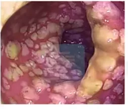
- The toxin released by C. difficile leads to the accumulation of a necrotic material which is responsible for the formation of a puss containing material (necrotic material) on the membrane of the intestine.
- ENDOSCOPIC VIEW : A pseudomembrane formation can be seen in the endoscopy, due to the formation of the pus containing material on the intestinal membrane. The presence of this pseudo membrane gives this disease its name.
- MICROSCOPIC VIEW:

Important information
- The presence of Necrotic debris and neutrophils arising from the epithelial lining of the intestine can be seen, forming a “Mushroom-like” appearance or “volcanic eruption” form in the microscopic view.
- TREATMENT:
- Vancomycin
- Fidaxomicin
- Metronidazole
Also Read: Inflammation Types - NEET PG Pathology
Trisomy 18: Causes, Symptoms, Types, Diagnosis, Treatment and Prevention
Frequently Asked Questions
Q: ‘Anchovy sauce” pus can be seen in which disease?
Answer: Amoebiasis
Q: What is the most common cause of Pseudomembranous colitis?
Answer: Excessive antibiotic use
Q: What is the treatment of Amoebiasis?
Answer: Antibiotics like metronidazole are used for the treatment of Amoebiasis.
Q: Which organism causes Intestinal tuberculosis?
Answer: Mycobacterium bovis.
To study intestinal ulcers and infections in detail, you can download the PrepLadder app and find in-depth video lectures on the topic. In addition to that, also find relevant study notes and MCQs for practice and strengthen your NEET PG exam preparations.

PrepLadder Medical
Get access to all the essential resources required to ace your medical exam Preparation. Stay updated with the latest news and developments in the medical exam, improve your Medical Exam preparation, and turn your dreams into a reality!
Navigate Quickly
Tuberculosis
Enteric fever/typhoid
Hints to look for
Amoebiasis
Psuedo membraneous colitis
Frequently Asked Questions
Top searching words
The most popular search terms used by aspirants
- Medical PG Pathology
- Medical PG Preparation
- NEET PG Pathology