Thalassemia - Symptoms, Causes, Indicatives and Diagnosis
Feb 7, 2023

Thalassemia is an inherited blood disorder which hampers the body’s ability to produce healthy red blood cells and hemoglobin.
This blogpost will cover the Thalassemia meaning, its types, symptoms and causes in detail which will further help you in your NEET-PG preparation. So, let’s get started with the symptoms of Thalassemia.

Thalassemia Symptoms
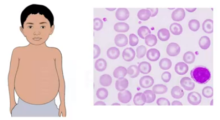
- Body abnormalities including an abnormal facial appearance, presence of pallor, presence of hepatosplenomegaly etc. are shown by individuals having thalassemia. Positive family history is most commonly the cause of thalassemia.
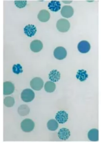
- PERIPHERAL SMEAR OBSERVATIONS:
- Microcytic hyperchromic RBCs can be seen.
- Cells having a “bull’s-eye” appearance can be seen. These are called the target cells.
- Beta-thalassemia is the most common type of thalassemia among the Indian population. Regular blood transfusion is required for people who are suffering from thalassemia major.
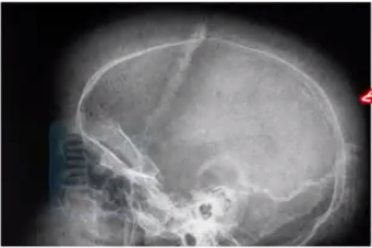
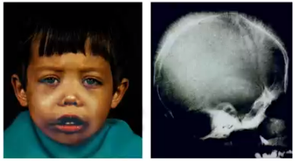
- Thalassemia intermedia patients only need occasional blood transfusion. Patients with mild thalassemia maintains Hb levels between 9 and 12 g/dl and do not require any specific treatment. The patients will be having no history of blood transfusion. Patients having thalassemia show manifestations such as extramedullary hematopoiesis. A hair on end appearance can be seen on the X-ray skull of the patients. Enlargement of the maxilla caused by bone marrow expansion results in a characteristic appearance known as chipmunk faces (depressed nose, prominent maxilla) in thalassemia patients.
Major Indicatives Of Thalassemia
- Dyspnea
- Pallor
- Stunted growth
- Chipmunk face
- Malocclusion of teeth
- Hair on end X-ray skull appearance.
Also Read:
Diagnosis of Thalassemia
Diagnosis is confirmed by carrying out globin gene sequencing, High-performing liquid chromatographic examination of the Hb, hemoglobin electrophoresis etc. In case of an individual having thalassemia trait, there is always a risk of inheriting the condition to their next generation.
- NESTROFT TEST (Naked Eye Single Tube Red Cell Osmotic Fragility Test):
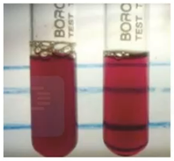
- It is a valuable and cost-effective thalassemia screening test. When the blood sample of a normal individual is put inside a hypertonic saline solution, hemolysis takes place, because of which the lines will be visible on the back of the test tube. This is seen as the control. When the same procedure is carried out for a test sample (blood sample of a patient having thalassemia trait), because of the reduced osmotic fragility, hemolysis doesn’t take place. As a result of this, the black line won’t be visible on the back of the test tube. NESTROFT Test is only an example of an indicator. Hence, it can only be considered as a screening test and not a confirmatory test.
Give a boost to your Pathology preparation with the INI-CET November 2022 recall questions by Dr Preeti Sharma, our faculty for Pathology.
Also Read: Cell Injury and Cell Dealth
Differences between Thalassemia Trait and Thalassemia Major
- When performing HPLC, in patients having thalassemia trait, an elevated concentration of HBA2 can be found. When the HBA2 percentage is more than 3.5, it is a good indicative of thalassemia trait. Whereas, in case of thalassemia major, also known as Mediterranean anemia or Cooley’s anemia, the predominant hemoglobin present in these individuals is HbF. The history of blood transfusions can also help to differentiate between thalassemia trait and thalassemia major.
- Alpha Thalassemia :
- It is the uncommon variant of thalassemia. Characterized by the presence of gene deletion. HbH precipitates within the RBCs can be found in patients with alpha thalassemia. This condition is known as the “golf ball” appearance.
Important Information
- Presence of microcytic hyperchromic cells and target cells can also be seen in the case of iron deficiency anemia.
- Iron deficiency anemia and thalassemia can be distinguished from each other by calculating the Mentzer index (obtained by dividing MCV with the RBC count). If the value turns out to be <13, it is very much a suggestive of thalassemia and if the value is >13, it is an indicative of iron deficiency anemia.
Also Read:
CHRONIC INFLAMMATION: Symptoms, Causes, Diagnosis, and Treatment: Pathology
Acute inflammation: Symptoms, Causes, and Treatment : Pathology
To scale up your NEET PG preparation with the best-in-class video lectures, QBank, Mock Tests and more, download the PrepLadder App!
Download PrepLadder's NEET PG app for Android
Download PrepLadder's NEET PG app for iOS

PrepLadder Medical
Get access to all the essential resources required to ace your medical exam Preparation. Stay updated with the latest news and developments in the medical exam, improve your Medical Exam preparation, and turn your dreams into a reality!
Navigate Quickly
Thalassemia Symptoms
Major Indicatives Of Thalassemia
Diagnosis of Thalassemia
Differences between Thalassemia Trait and Thalassemia Major
Top searching words
The most popular search terms used by aspirants
- Medical PG Preparation
- NEET PG Preparation