Formation of the Germ Layers : Gastrulation, Migration of Primordial Germ Cells
Jun 19, 2023

During the development, the three germ layers, which are known as ectoderm, endoderm and mesoderm are formed by the same epiblast cells and are formed at the third weekend this process is known as Gastrulation. Epiblast cells undergo proliferation that leads to the formation of a Primitive streak. Epiblast cells are migrating to the ventral side to replace the hypoblast cells, they become endoderm.
Some of the epiblast cells will form the mesoderm. The remaining epiblast cells will form an ectoderm. In the later stages, ectoderm will form the neural tube, mesoderm will form the cardiovascular tube and endoderm will form the gut tube. There is a cephalocaudal folding which means the head comes towards the tail.
Read this blog further to get a quick overview of this important topic for anatomy and ace your NEET PG exam preparation.
Gastrulation
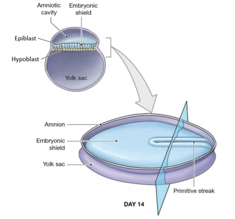
It is the process of formation of three germ layers at the third week of the development. Mast cells are proliferating at the floor of the amniotic cavity to form primitive streaks. In the dorsal view of the conceptus, all these epiblasts form a disc-shaped structure.
On Days 14 - 15, thickening will appear on the disc which is known as the Prechordal plate. Thickening is before the notochord formation. Wherever the prechordal plate is presented that will become the cephalic/anterior end of the baby, the Opposite end is the caudal end of the baby. The primitive streak will appear towards the tail (caudal end of the baby). It migrates towards the head region (cephalic end).
Primitive streak which is the proliferation of epiblast appears in the caudal end and moves towards the cephalic region known as the Caudocephalic region. A gene is required here called a homeobox gene (HOX) is going to destroy the cephalocaudal end of the baby.
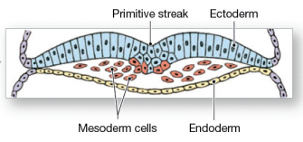
All this will happen at the floor of the amniotic cavity. This primitive streak will appear on day 14 (end of week 2). At the floor of the amniotic cavity, the epiblast cells form the primitive streak. These cells will migrate into the primitive grooves towards the ventral side and reach the roof of the yolk sac cavity. Epiblast cells will form the first germ layer endoderm and form the middle layer, mesoderm. The remaining epiblast cells in the dorsal side will become ectoderm.
Three germinal layers
- First: Endoderm
- Second: Mesoderm
- Third: Ectoderm
- Ectoderm germ layer derived from epiblast will contribute to neural tube
- Mesoderm contributes to cardiovascular tube
- Endoderm contributes to the gut tube

- Neural tube forming CNS which includes the brain towards the head and tail towards the spinal cord
- Cardiovascular tube location keeps changing along the folding of the embryo
- The gut tube will further from the 3 components:
- Foregut
- Midgut
- Diverticulum: Vitelline duct entering into the umbilical cord
- Hindgut
- Diverticulum: Allantois entering into the umbilical cord
Development during week 3 & 4
- Week 1: Blastocyst (blast cells and a cyst-like cavity)
- Week 2: Blastocyst
The above two stages are called pre-embryonic stages
- Week 3: Embryo (Conceptus as an embryo)
Blastocyst has blast cells and a cyst-like cavity that are called the inner cell mass (embryoblast). The outer cell mass is made of trophoblasts, they secrete nutrition for the baby. In 6 to 8 days, the trophoblasts will undergo division and lose their cell boundaries to form a syncytium ( multinucleated) called syncytiotrophoblast.
As these cells are intact in the cell membrane, they are called cytotrophoblasts. In these 6 to 8 days, other cells also divide into two (embryo blasts that form the embryo). These embryo blasts are divided into
- Dorsal epiblast (floor of the amniotic cavity)-Cells are columnar
- Ventral hypoblast (at the roof of the yolk sac cavity)-Cuboidal cells
Week two means week of two (the two cavities, two embryo blasts, two trophoblasts). By the end of week two (14-15), a primitive streak will be formed (proliferation of epiblast cells). Some of the cells are isolated from the epiblast by the end of the second week called primordial germ cells (derived from epiblast).
In Week 3 Epiblast cells will migrate towards the ventral side, to display the hypoblast cells to take their space for becoming endoderm (first germ layer). These hypoblasts cells are removed by the epiblasts, they will be lining the yolk sac cavity (forming extra embryonic endoderm). Embryonic endoderm is formed by the epiblast cells.
Middle layer formed is mesoderm and the outer layer is ectoderm.Epiblast forms some of the cells in the ectoderm region known as NCC cells. These neural crest cells are derived from epiblasts (fourth germ layer). Ectoderm changes into two types:
- Centrally developing neural ectoderm
- Ectoderm at the periphery: Surface ectoderm
Primordial germ cells have been isolated from the primitive streak as early as Day 14-15. Earliest derivative epiblast cell is primordial germ cell
Q. Which of the following is NOT TRUE regarding gastrulation
- Occurs at 3rd week
- Epiblast cells on inner mass form all germ layers
- Establishes all the three germ layers
- Occur at the caudal end of the embryo prior to its cephalic end
Answer: Occur at the caudal end of the embryo prior to its cephalic end.
Related Anatomy Articles :
Migration of Primordial Germ Cells
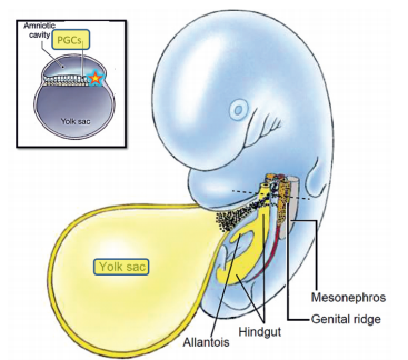
Primordial germ cells are the first sex cell of the body. PGCs are isolated from the epiblast's cell at the primitive streak by the end of week 2. PGC is the first derivative of epiblast in temporal sequence by Day 14-15. Later they migrate towards the endodermal wall of the yolk sac.
Earlierly, they used to think that primordial germ cells developed from the endodermal wall of the yolk sac but now it has been realized that they came from the epiblasts.
At week 4 these cells migrate from the yolk sac towards the genital ridge using dorsal mesentery of hindgut. The primordial cells should migrate to the genital ridge of the abdomen to form gametes (gametogenesis).
At the end of week 5, primordial cells will reach the genital ridge. Oropharyngeal teratoma may occur if the primordial cells move towards the oropharynx. If primordial germ cells is going towards the tail of the embryo, sacrococcygeal teratoma, Abrupt migration of primordial germ cells may cause oropharyngeal teratoma and sacrococcygeal teratoma. Longitudinal section of embryo
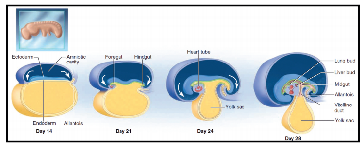
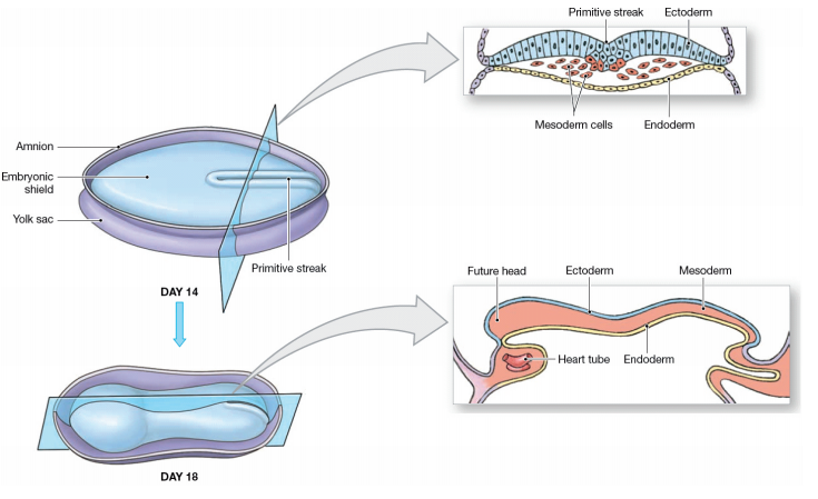
Week 3 & 4, head end of baby fold ventrally and tail end coming close to each other, the dorsal cavity (amniotic cavity) is becoming large and surrounding the baby whereas the ventral cavity becoming narrow and regresses and disappear.
Yolk Sac and Amniotic Cavity

Amniotic cavity becomes large and almost covers the embryo caudally as well as the cephalic region. Yolk sac cavity being incorporated into the embryo basically the gut tube all lined by endoderm. Gut tube is Foregut, Midgut, Hindgut. Vitelline duct and Allantois regresses and become the content of umbilical cord. Neuro ectoderm develops CNS (brain and spinal cord). Mesoderm derives cardiovascular tube.
.jpg)
Transverse Section of the Embryo
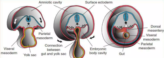
Dorsal amniotic cavity folding to the right as well as left side, Covers most of the embryo. Ventral yolk sac cavity becomes continuous with the gut tube and only the gut tube will persist and the yolk sac cavity will disappear. The 3rd cavity is developed which is known as the Intraembryonic coelomic cavity. Coelomic cavity develops within the embryo. Later, the coelomic cavity develops into 3 parts
- Pericardial cavity
- Pleural cavity
- Peritoneal cavity- Peritoneal cavity lined by peritoneum. Double fold peritoneum is suspending the gut tube from the dorsal side known as Dorsal mesentery.
- The ventral yolk sac cavity become continuous with the gut tube and only the gut tube will persisting and yolk sac cavity will disappear leaving behind endodermal gut tube.
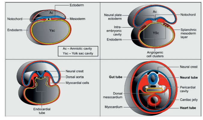
- Neural ectoderm forming neural plate → Neural groove → Neural tube → CNS.
- Lateral plate Mesoderm → Splanchnic mesoderm layer → Myocardial cells → Myocardium or Cardiovascular tube. Heart tube will be suspended with the help of the dorsal mesocardium.
To scale up your NEET PG preparation with the best-in-class video lectures, QBank, Mock Tests and more, download the PrepLadder App!
Download PrepLadder's NEET PG app for Android
Download PrepLadder's NEET PG preparation for iOS

PrepLadder Medical
Get access to all the essential resources required to ace your medical exam Preparation. Stay updated with the latest news and developments in the medical exam, improve your Medical Exam preparation, and turn your dreams into a reality!
Navigate Quickly
Gastrulation
Three germinal layers
Development during week 3 & 4
Migration of Primordial Germ Cells
Yolk Sac and Amniotic Cavity
Transverse Section of the Embryo
Top searching words
The most popular search terms used by aspirants
- NEET PG Anatomy
PrepLadder Version X for NEET PG
Avail 24-Hr Free Trial